
|
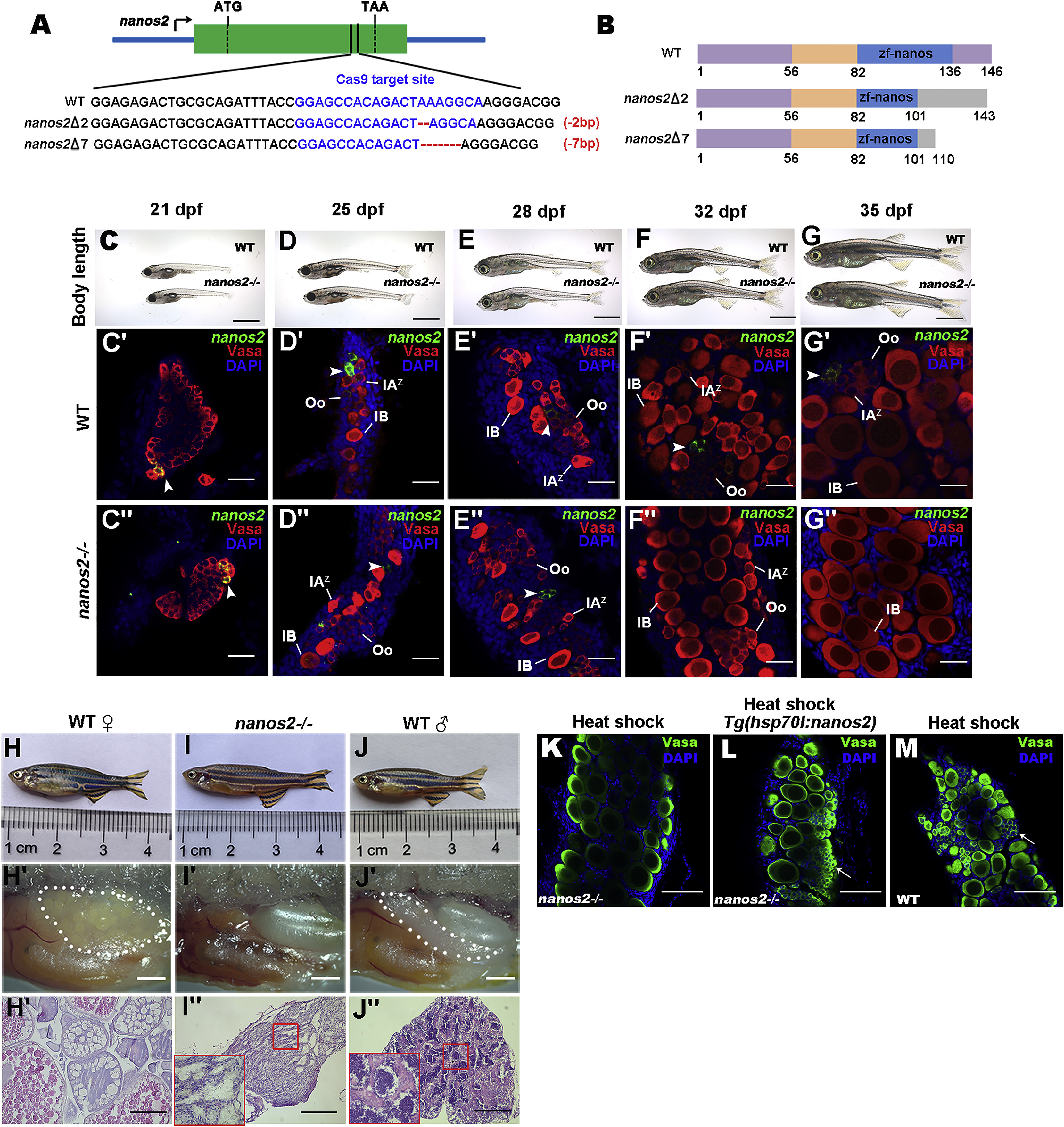
Fig. 1
nanos2 Is Required for Maintaining GSCs
(A) Schematic diagram of nanos2 genomic structure and mutation genotypes.
(B) Schematic representation of the protein functional domains of nanos2 from wild-type (WT) and two kinds of mutants (Zf-nanos, Zinc finger-nanos).
(C–G) Body length of wild-type (WT) and nanos2homozygous at 21 (C), 25 (D), 28 (E), 32 (F), and 35 (G) dpf.
(C′–G″) Triple fluorescent labeling with FISH-nanos2, anti-Vasa antibodies, and 4′,6-diamidino-2-phenylindole (DAPI) staining in the ovaries of wild-type and nanos2 homozygous at 21, 25, 28, 32, and 35 dpf. Note loss of GSCs (arrowheads) in the mutant starting at 32 dpf.
(H–J) Adult wild-type female (H), the nanos2 mutant (I), and wild-type males (J).
(H′–J′) Lateral views of gonads.
(H″–J″) H&E staining of gonad sections shows that all of the nanos2 mutants develop into the sterile males.
(K–M) At 40 dpf after heat-shock, double staining by anti-Vasa antibodies, and DAPI in the nanos2 mutants with (L) or without (K) Tg(hsp701:nanos2) transgenic background, and in the wild-type (M) shows that overexpression of nanos2 could rescue the phenotypes of nanos2 mutant. Arrows indicate early germ cells.
Oo, oogonia; IAz, zygotene-stage-IA; IB, stage IB oocyte. Scale bars: 500 μm (C–G), 50 μm (C′–G′′), 1 mm (H′–J′), 200 μm (H′′–J′′), 50 μm (K–M). See also Figure S1.