
Image
|
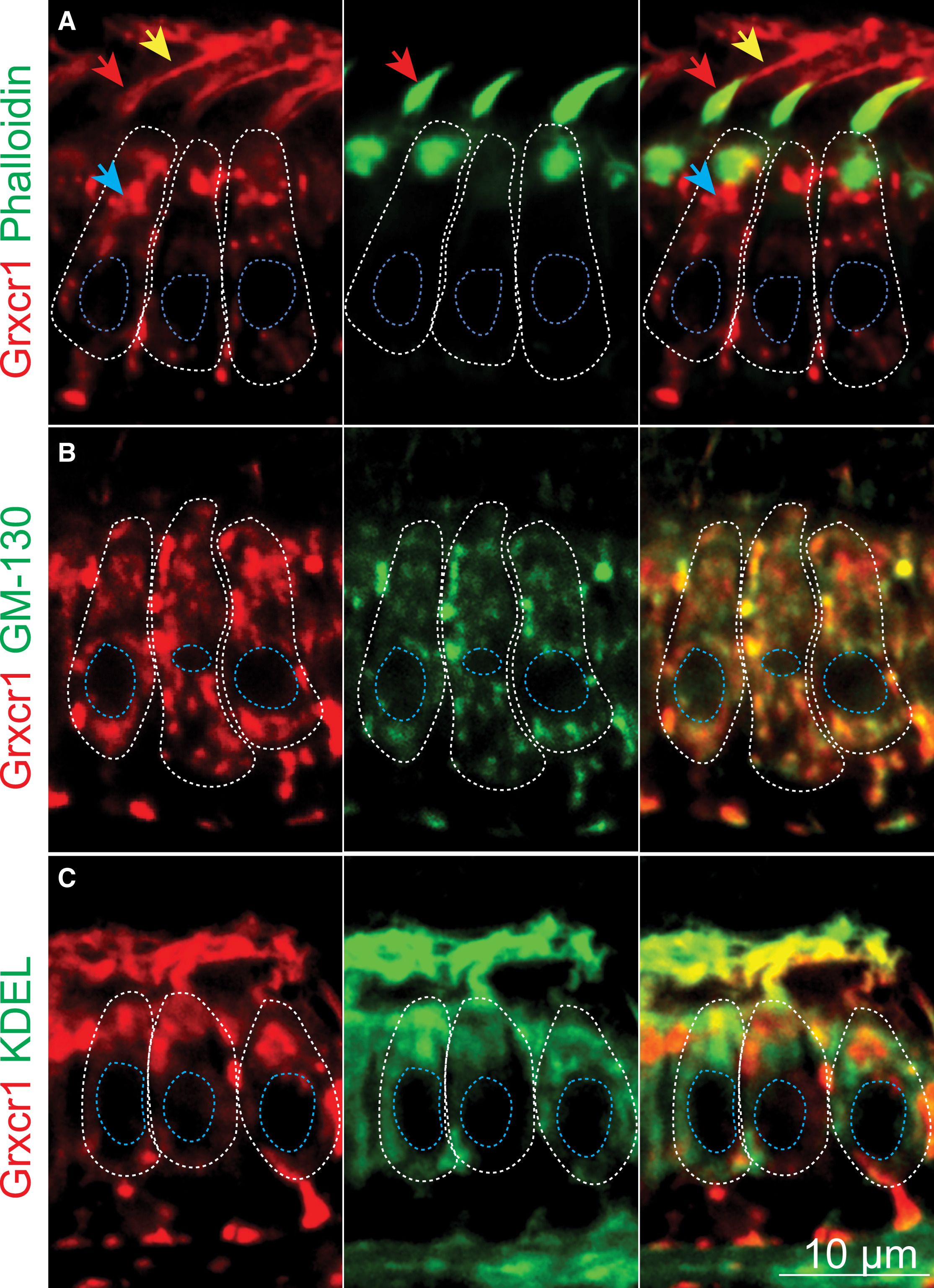
Figure Caption
Fig. 5
Grxcr1 Localizes in the Golgi Apparatus and ERGIC of the Hair Cells
(A–C) Confocal sections of the anterior macula. Shown are partial colocalization of Grxcr1 (red) and phalloidin (A), GM130 (B), or KDEL (green, C). Individual hair cells are outlined in white. Blue, red, and yellow arrows show Grxcr1 localization in the cell body, hair bundles, and kinocilia, respectively. The confocal sections were selected based on the localization of the organelles of interest (hair bundles, Golgi, and ER), hence the differences in Grxcr1 localization. Nuclei are outlined in blue; 5 dpf.
Figure Data
Acknowledgments
This image is the copyrighted work of the attributed author or publisher, and
ZFIN has permission only to display this image to its users.
Additional permissions should be obtained from the applicable author or publisher of the image.
Full text @ Cell Rep.