
Fig. 9
- ID
- ZDB-IMAGE-180823-11
- Genes
- Antibodies
- Publication
- Moreno et al., 2018 - Investigation of Islet2a function in zebrafish embryos: Mutants and morphants differ in morphologic phenotypes and gene expression
- All Figures
- Figures for Moreno et al., 2018
|
Fig. 9
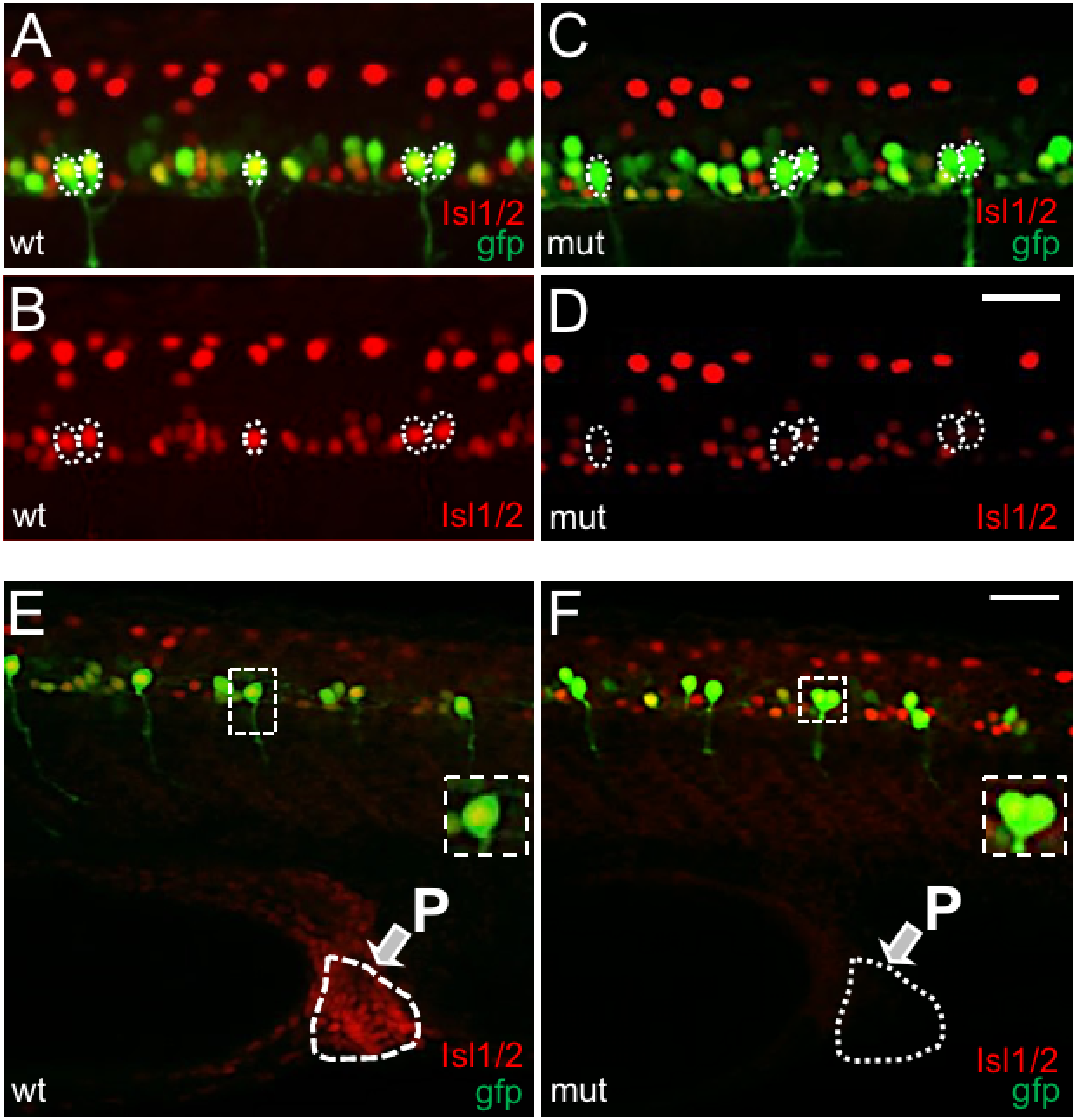
CaPs expressed reduced levels of Isl1/2 immunoreactivity in islet2a mutant embryos.
(A) In 28 hpf tg(mnx1:gfp) wildtype (wt, n = 15) embryos, CaPs (*) expressed both gfp (green) and Isl1/2 immunoreactivity (red), as revealed by the merged yellow signal. (B) The Isl11/2 immunosignal of Panel A is shown separately. In A-D, dotted lines circle the cell bodies of CaP/VaPs that were immunopositive for Isl1/2. In comparison to other ventral neurons, CaP Isl1/2 immunolabeling was more intense. (C) In 28 hpf tg(mnx1:gfp) mutant (mut, n = 14) embryos, CaPs (*) expressed gfp. However, compared to wildtype (A), the CaP Isl1/2 fluorescent immunolabel signal was less intense. Further, other ventral neurons continued to display Isl1/2 immunoreactivity. (D) The Isl1/2 signal of Panel C is viewed separately. Compared to wildtype (B), CaPs expressed reduced levels of Isl1/2 immunoreactivity. Further, despite the weak signal in CaPs, Isl1/2 immunoreactivity was present in other ventral neurons at levels similar to wildtype (B). Scale bar in D for A-D: 25 μm. (E, F) Examination of CaP Isl1/2 immunolabeling dorsal to the proctodeum. (E) In wildtype embryos, both the proctodeum (white arrow) and CaPs (asterisks) displayed Isl1/2 immunolabeling. One CaP, contained within white dotted line box, is shown at higher magnification in the inset. (F) In mutant embryos, Isl1/2 immunolabeling was not detected in the proctodeum, consistent with loss of Islet2a protein expression. Despite this, a low level of Isl1/2 immunoreactivity persisted in CaPs (asterisks; one CaP shown at higher power in inset).