
Fig. 2
|
Fig. 2
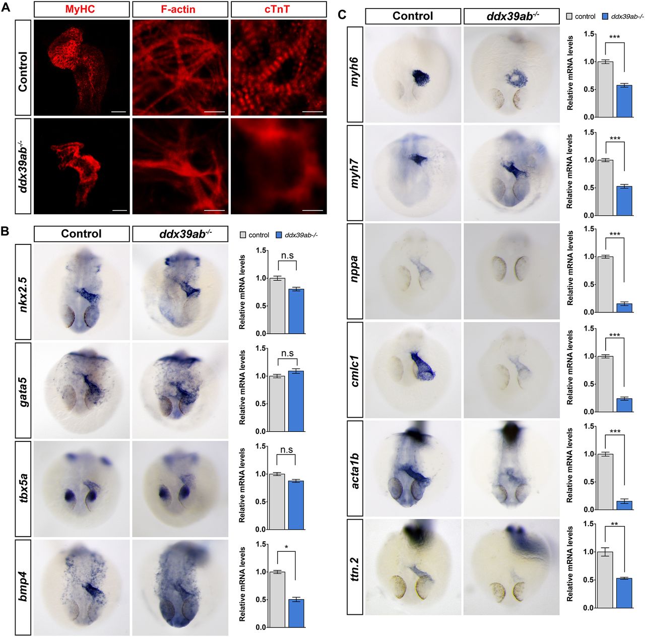
Loss of ddx39ab leads to cardiomyocyte differentiation defects in zebrafish embryos. (A) Ventral view of heart from whole-mount wild-type or ddx39ab mutant zebrafish embryos at 36 hpf, with cranial to the top. Myocardium was labeled with MF20 antibody. Scale bars: 200 μm. (B) RNA in situ hybridization and qPCR results for cardiogenic regulatory gene expression in wild-type and ddx39ab mutant zebrafish embryos at 26 hpf. (C) RNA in situ hybridization and qPCR results for cardiomyocyte structural gene expression in wild-type and ddx39ab mutant zebrafish embryos at 26 hpf. (B,C) Frontal views with dorsal side to the top. At least 15 (A) or 20 (B,C) embryos for each genotype were analyzed and representative samples are shown. For qPCR results, data are mean±s.e.m. n.s, not significant. *P<0.05, **P<0.01, ***P<0.001.