
|
Fig. 6
Dynamics of Germline Reprogramming in Live Zebrafish Embryos
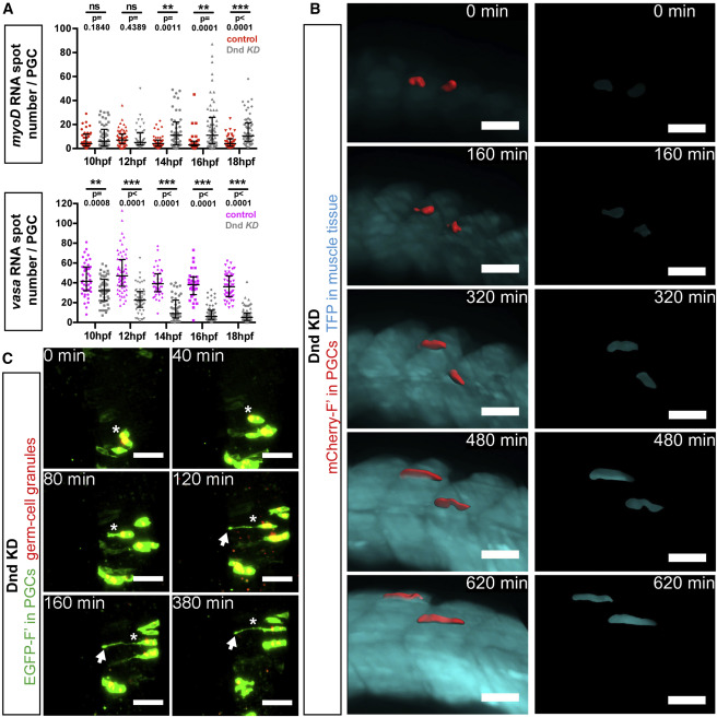
(A) Quantification of myoD-positive (top) and vasa-positive (bottom) RNA spots in 10–18 hpf cxcl12a−/− embryos. Number of RNA spots in PGCs of embryos knocked down for Dnd are represented by gray dots (upper and bottom panels), showing an increase in myoD RNA spots (red dots, upper panel) and a decrease in vasa RNA spots (pink dots, bottom panel), relative to control PGCs. p Values were determined using the Mann-Whitney U test with α = 0.05. 31 ≤ n ≤ 65 (n, number of cells). Data are presented as median ± IQR.
(B) PGCs (segmented, red) located within the developing muscle tissue (blue) in live Dnd-deficient embryos knocked down for Cxcl12a (left panels). Panels on the right show expression of the muscle-specific reporter within the segmented PGCs. Light sheet microscopy images were captured between 14 and 24 hpf and are presented as 3D projections. Scale bars, 50 μm.
(C) PGCs (green) located within the region of the developing neural tube in Dnd-deficient embryos knocked down for Cxcl12a. Germ cell granules were visualized using a DsRed-Granulito protein fusion. Asterisks mark a single PGC undergoing reprogramming to become a neuron-like cell. Arrows point to an axon-like protrusion. Light sheet microscopy images were acquired in 12- to 19-hpf embryos. Scale bars, 50 μm.
F′, farnesylated; KD, knockdown; hpf, hours post fertilization. See also Figure S4 and Movies S5, S6, and S7.
Reprinted from Developmental Cell, 43, Gross-Thebing, T., Yigit, S., Pfeiffer, J., Reichman-Fried, M., Bandemer, J., Ruckert, C., Rathmer, C., Goudarzi, M., Stehling, M., Tarbashevich, K., Seggewiss, J., Raz, E., The Vertebrate Protein Dead End Maintains Primordial Germ Cell Fate by Inhibiting Somatic Differentiation, 704-715.e5, Copyright (2017) with permission from Elsevier. Full text @ Dev. Cell