
|
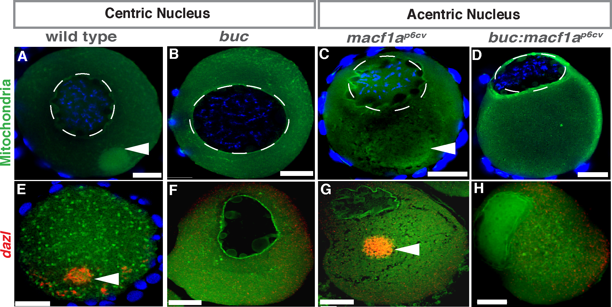
Fig. 2
Epistasis of bucky ball and macf1a in nuclear positioning.
A-D) DiOC6 staining (mitochondria, green) and dazl in situ (E-H, red) in stage I oocytes label the Bb. (A, E) WT with centered nucleus and Bb present. (B, F) buc mutant with centered nucleus, absent Bb, and unlocalized dazl. (C, G) macf1a mutant with acentric nucleus and Bb enlarged. (D, H) macf1ap6cv; buc double mutant with acentric nucleus, absent Bb, and unlocalized dazl. DAPI (blue) stains DNA (A-D). DiOC6: N≥ 3 ovaries; >30 WT, >30 macf1ap6cv, 15 bucp106re, 24 macf1ap6cv; bucp106re oocytes. dazl in situ: N = 3 ovaries; 10 bucp106re, 7 macf1ap6cv, 14 macf1ap6cv; bucp106re oocytes. Representative images from 2 experiments. Dotted white lines outline the nucleus. Images are a sum of 3 single optical confocal sections. Arrowheads indicate the Bb. Scale bar: 20 μm.