
|
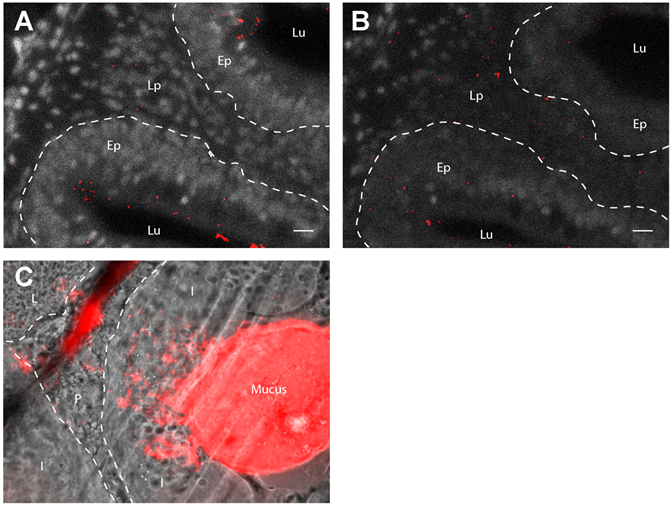
Fig. S2
Artefactual nanoparticle displacement at cryosection surface. (A, B): Images of 40µm-thick gut cryosections from adult zebrafish exposed for 24h to 0.05% red fluorescent nanoparticles. Pictures are maximal intensity projection of 5 confocal pictures taken every two micrometers. Whereas acquisitions in the core of the cryosection reveal low nanoparticle uptake in the lamina propria (A), a larger number of nanoparticles are detected in the same region at the surface the cryosection (B), suggesting that PLA nanoparticles may be displaced during immersion steps of the staining protocol. (C): Epifluorescence image from similar cryosections, cut across a nanoparticle-rich region of the gut lumen. The smear of nanoparticles originating from the lumen suggests that the cryosecting blade may displace nanoparticles at the surface of sections. Lu (Lumen), Lp (Lamina propria), Ep (Epithelium), I (Intestines), P (Pancreas), L (Liver). Scale bar: 10 µm