
|
Fig. S2
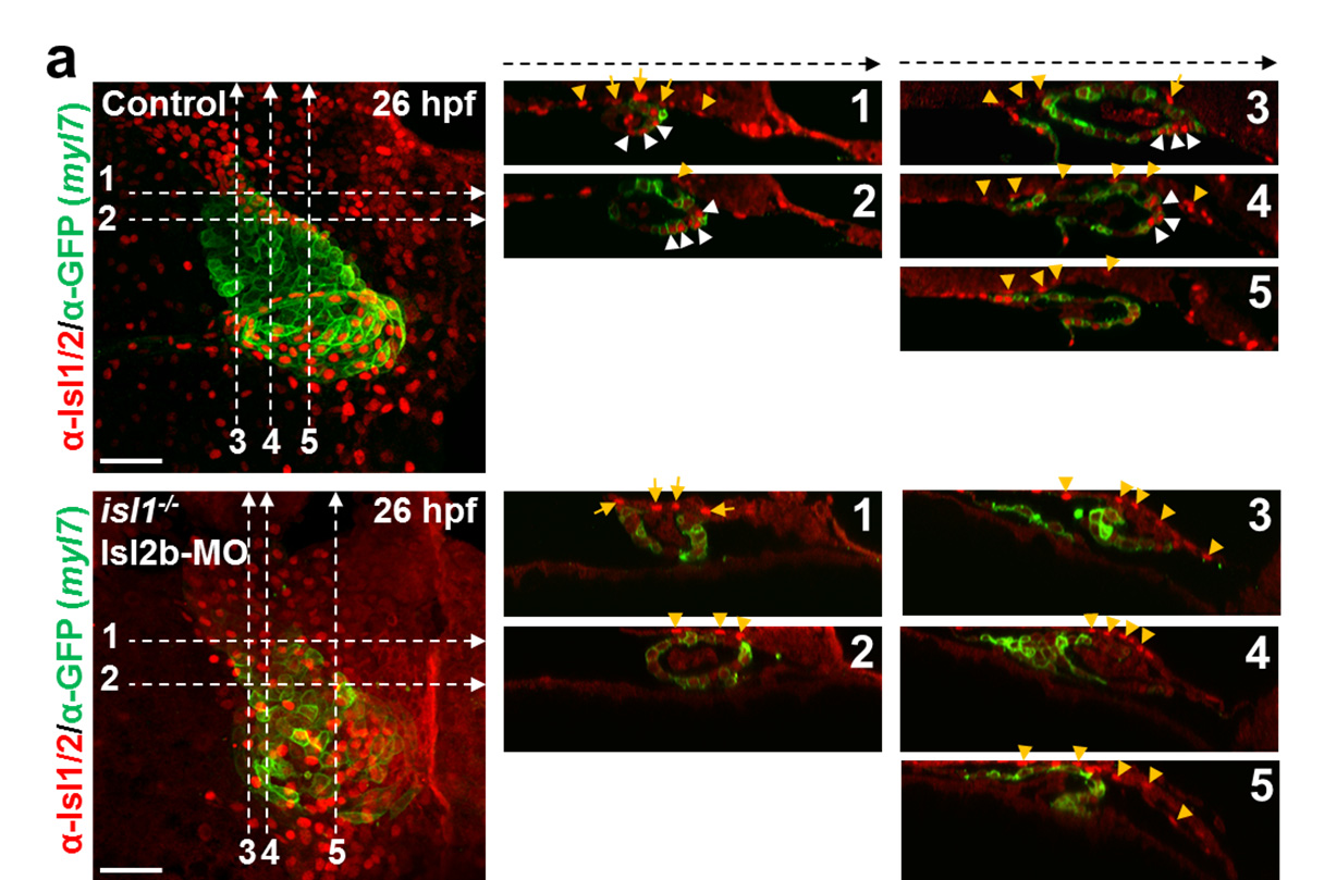
Distinct patterns of expression of Islet family members in the developing heart.
(a) Confocal images of control and Isl2b morpholino-injected Tg(myl7:EGFP-HsHRAS)s883 isl1-/- embryos stained with anti-GFP and anti-Isl1/2 antibodies at 26hpf. Optical sections showing residual Isl2a+ cells in the pericardial wall adjacent to the arterial pole (orange arrows) and in endodermal cells on top of the heart tube (orange arrowheads) in Isl2b morpholino-injected isl1- /- embryos. Isl2b+myl7+ expressing cells (white arrowheads in wild-type embryos) are lost in Isl2b morpholino-injected isl1-/- embryos. (b) Confocal images of control and Tg(kdrl:EGFP)s843isl1-/- Isl2b morpholino-injected embryos stained with anti-GFP and anti-Isl1/2 antibodies at 26 hpf. Optical sections showing Isl1+flk1+ cells located in the endocardium of the forming ventricle, as well as in the vessels at the arterial pole (blue arrowheads) in wild-type embryos. These cells are not detectable in isl1-/- Isl2b-MO embryos, suggesting that they do not express Isl2a. Scale bars, 50 μm.