
Fig. 4
|
Fig. 4
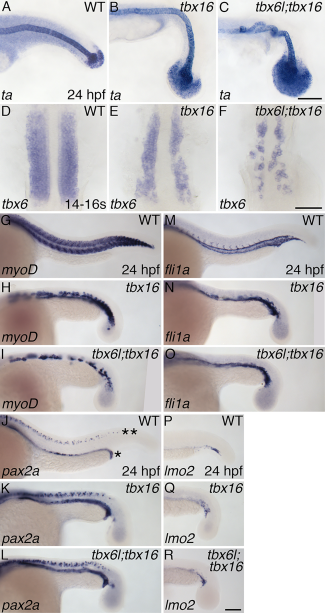
tbx6l and tbx16 have partially redundant roles in formation of tail paraxial mesoderm. A–R: In situ hybridization for ta (A–C), tbx6 (fss) (D–F), myoD (G–I), pax2a (J–L), fli1a (M–O), and lmo2 (P–R) at 24 hpf (A–C;G–R) and the 14- to 16-somite stage (D–F). Not shown are tbx6l single-mutant embryos as they are indistinguishable from wild-type embryos. In panel J, the row of pax2a-expressing pronephros cells is indicated by an asterisk (*), and the row of pax2a-expressing spinal cord neurons is indicated by a double asterisk (**). Scale bars in C (for A–C) and R (for G–R) = 100 μm. Scale bar in F (for D–F) = 50 μm.