
|
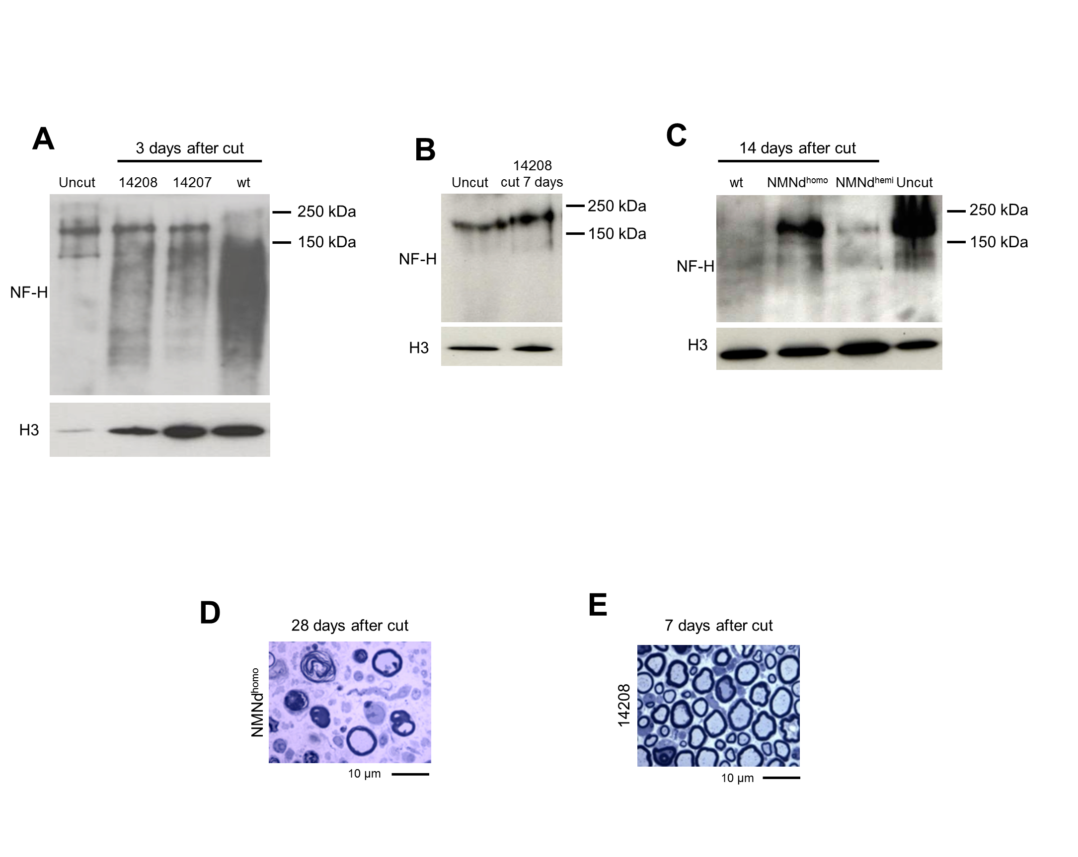
Fig. S1
Protection of injured axons in NMN deamidase transgenic mice, related to Figure 3.
(A) Western blot showing the extent of NF-H degradation in distal tibial nerve of founder 14207 and 14208 (NMNdhemi) mice 3 days after cut (relative to uncut and cut wild-type (wt) nerves).
(B) Western blot showing NF-H degradation 7 days after cut in a founder 14208-derived NMNdhemi mouse (sterile offspring of founder 14208).
(C) Comparison of NF-H degradation in NMNdhemi and NMNdhomo mice (founder 14209-derived line) at 2 weeks after cut. In all the blots, histone H3 was used as a loading control.
(D) Light microscopy images of sciatic nerves of an NMNdhomo mouse (founder 14209-derived line) 28 days after axotomy and
(E) a founder 14208-derived NMNdhemi mouse 7 days after cut. Of note, protection at 7 days in the 14208-derived NMNdhemi mouse appears stronger than that observed in line 14209-derived NMNdhemi mice (Figure 3C). This is consistent with the higher enzymatic activity detected in tissue from founder 14208 compared to line 14209.