
Image
|
Figure Caption
Fig. S4
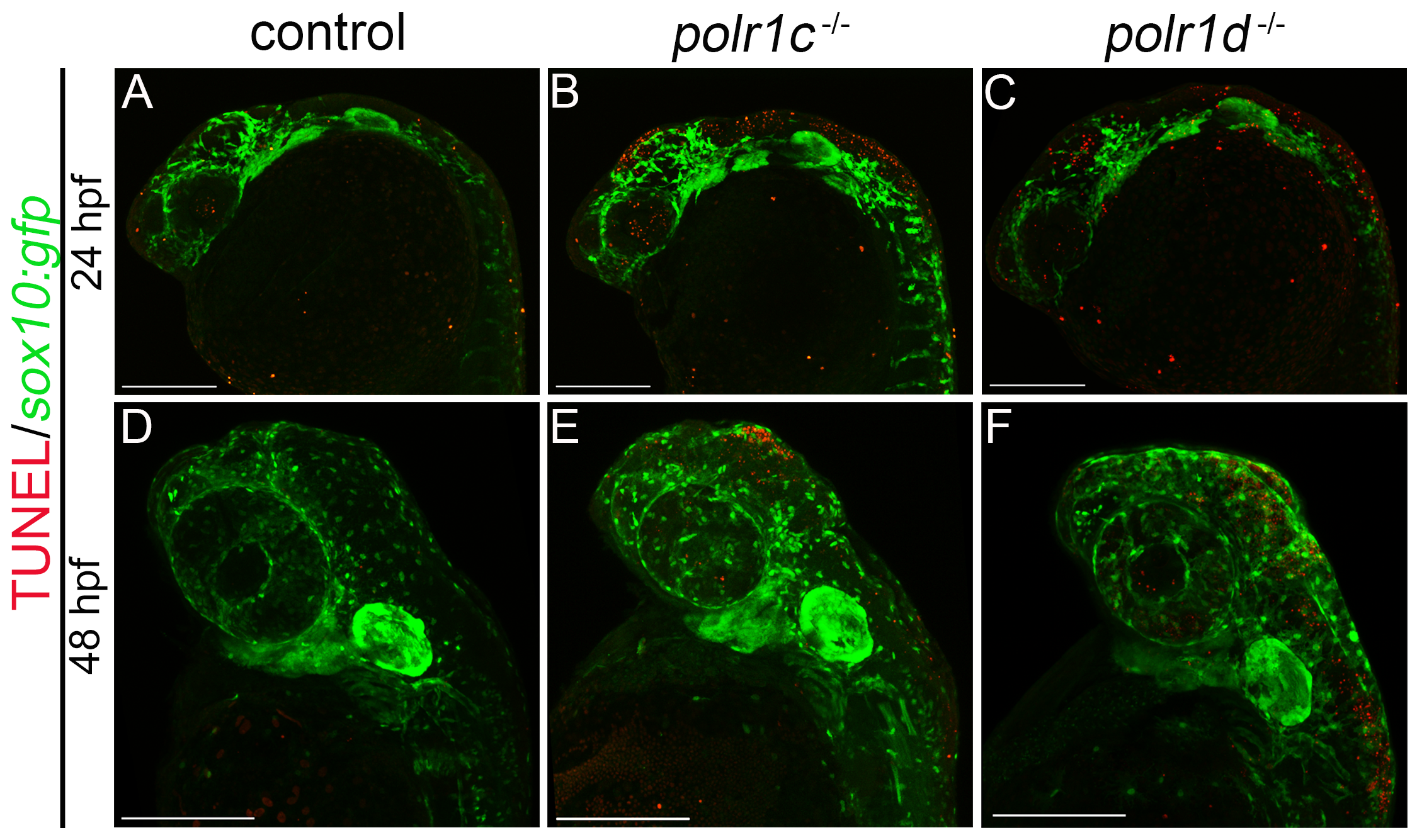
The migratory NCC population is not undergoing apoptosis in polr1c and polr1d mutant embryos.
(A-D) TUNEL staining in sox10:gfp embryos shows increased cell death in mutant embryos which does not co-localize with the migratory NCC population. At 24 hpf, increased cell death can be seen in the neuroepithelial region of polr1c-/- and polr1d-/- embryos. (E-F) At 48 hpf, increased cell death can be observed in regions of the brain and eye of mutant embryos, but not within the pharyngeal arches. Scale bar = 200 μm.
Figure Data
Acknowledgments
This image is the copyrighted work of the attributed author or publisher, and
ZFIN has permission only to display this image to its users.
Additional permissions should be obtained from the applicable author or publisher of the image.
Full text @ PLoS Genet.