
|
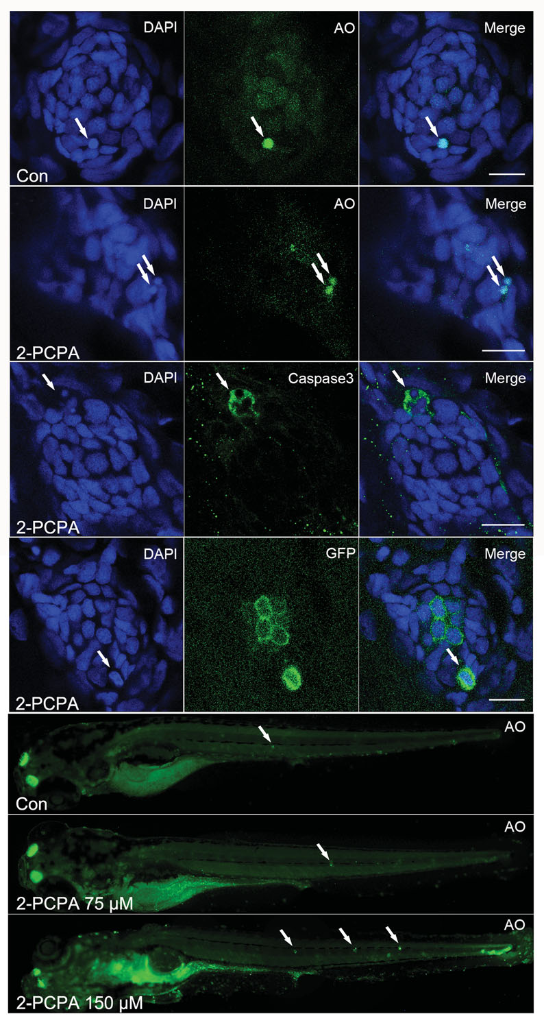
Fig. 4
High concentration of 2-PCPA treatment leads to apoptosis. (A-E) 2-PCPA treatment at high concentration (150 µM) induced an enhancement of cellular apoptosis within neuromasts. A-B: AO staining in the neuromast of a control (A) and a 2-PCPA treated larva (B) at 5 dpf. White arrows indicated AOpositive cells. (C) Cleaved Caspase-3 staining in the neuromast of a 150 µM 2-PCPA treated larva at 5 dpf. (D) The detection of hair cell death in neuromast. Neuromast images from brn3c:mGFP larvae treated with 150 µM 2-PCPA. White arrows in (D) showed dying hair cells being extruded from the neuromast. Scale bar =10 µm. (E) 150 µM 2-PCPA treated larva (lower panel) have high levels of acridine orange (AO) staining (white arrows) in neuromasts, compared with control (upper panel) and 75 µM 2-PCPA treated larva (middle panel) at 5 dpf.