
|
Fig. 6
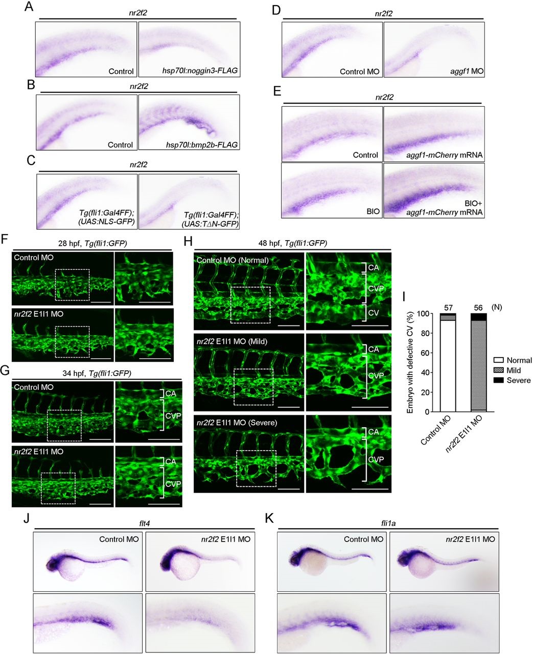
Bmp regulates CV formation by inducing Nr2f2 expression via β-catenin. (A-E) Expression patterns of nr2f2 mRNA in the caudal regions of 48hpf embryos. (A) Embryos injected without (control) or with hsp70l:noggin3-FLAG plasmid were heat shocked at 24hpf. (B) Embryos injected without (control) or with hsp70l:bmp2b-FLAG plasmid were heat shocked at 24hpf. (C) Tg(fli1:Gal4FF);(UAS:NLS-GFP) and Tg(fli1:Gal4FF);(UAS:TΔN-GFP) embryos. (D) Embryos injected with control MO or aggf1 MO. (E) Control embryos (top left) were injected with aggf1-mCherry mRNA, or incubated with BIO (a glycogen synthase kinase 3 inhibitor), or injected with aggf1-mCherry mRNA and then treated with BIO. (F-H) Confocal stack images of the caudal regions of Tg(fli1:GFP) embryos injected with control MO or nr2f2 E1I1 MO at 28 (F), 34 (G) and 48 (H) hpf. The boxed areas are enlarged to the right. Note that knockdown of Nr2f2 caused a variety of impairments in CV formation. Embryos with the mild phenotype lacked the CV, but developed the CVP (middle row in H), whereas those with severe phenotypes lacked the CV and exhibited defective CVP (bottom row in H). (I) CV phenotypes observed in F were quantified, as in Fig. 2D. (J,K) Expression patterns of flt4 (J) and fli1a (K) mRNAs in 36hpf embryos injected with control MO or nr2f2 E1I1 MO. Regions from the yolk tube to the tail are enlarged beneath. Scale bars: 100µm.