
|
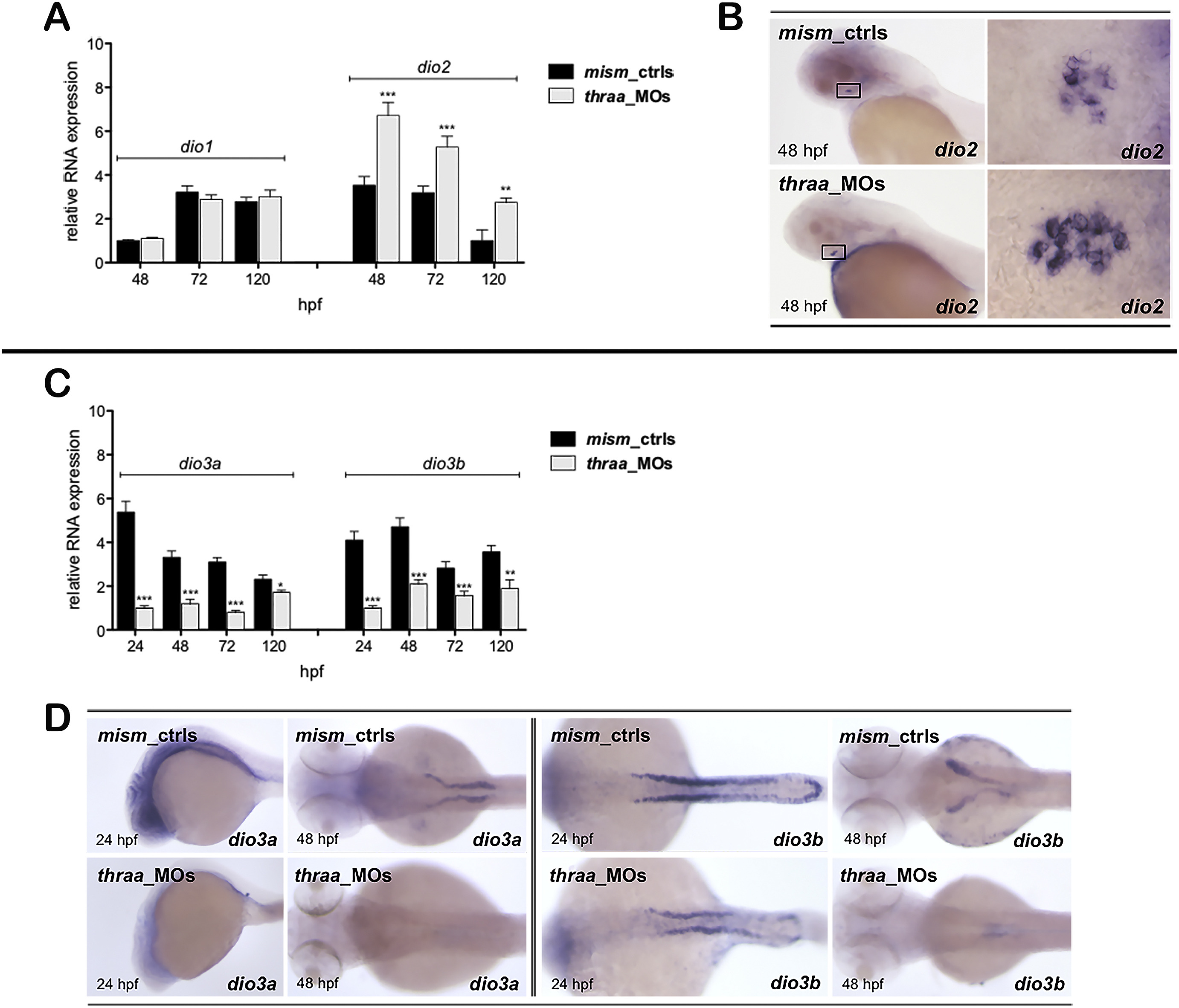
Fig. 8
Panels A and C) qRT-PCR of dio1, dio2, dio3a and dio3b expression in mism_ctrls and thraa_MOs at 24-120 hpf. *p < 0.05, **p < 0.01 and ***p < 0.001 vs mism_ctrls. Panel B) WISH of dio2 mRNA expression at 48 hpf: the pituitary dio2-positive cells are visible in lateral view, and in ventral view in the magnification of the corresponding square. Panel D) WISH of dio3a and dio3b mRNAs expression: dio3a is detectable in brain at 24 hpf (lateral view) and pronephric tubules at 48 hpf (dorsal view). The dio3b is also visible in pronephros at 24 and 48 hpf. The images are representative of 3 experiments (30 embryos each).
Reprinted from Molecular and Cellular Endocrinology, 424, Marelli, F., Carra, S., Agostini, M., Cotelli, F., Peeters, R., Chatterjee, K., Persani, L., Patterns of thyroid hormone receptor expression in zebrafish and generation of a novel model of resistance to thyroid hormone action, 102-17, Copyright (2016) with permission from Elsevier. Full text @ Mol. Cell. Endocrinol.