
|
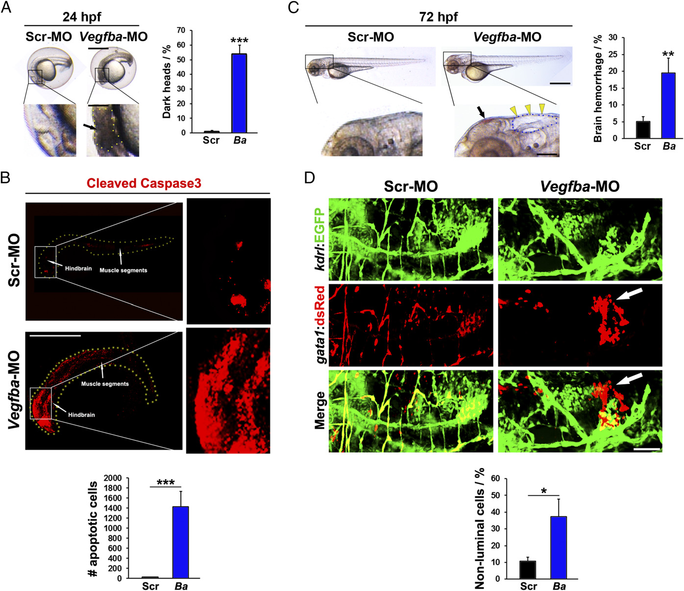
Fig. 2
Brain cell death and hemorrhage of VEGF-B–deficient zebrafish embryos. (A) Micrographs of embryos injected with 0.6 pmol Scrambled or Vegfba morpholino; embryos were imaged at 24 hpf. (Scale bar, 200 µm; bar in amplified picture, 50 µm.) Black arrow points to the brain area with dead cells, and dotted line encircles the dead area. Quantification of the percentage of embryos with obvious dead areas in the brain at 24 hpf (average n = 229 embryos/group). ***P < 0.001. (B) Confocal micrographs of embryos injected with 0.6 pmol Scrambled or Vegfba morpholino and stained for cleaved caspase3 activity at 24 hpf. (Scale bar, 500 µm.) Quantification of numbers of cleaved caspase3-positive cells (n = 18–21 embryos/group). ***P < 0.001. (C) Micrographs of embryos injected with 0.6 pmol Scrambled or Vegfba morpholino; embryos were imaged at 72 hpf. (Scale bar, 200 µm; in amplified picture, 50 µm.) Yellow arrowheads point to cerebral edema, and dotted line encircles the edema area. Black arrow points to brain hemorrhage. Quantification of the percentage of embryos with brain hemorrhages at 72 hpf (n = 222–295 embryos/group). **P < 0.01. (D) Confocal micrographs of Tg(Kdrl:egfp;Gata1:DsRed) embryos injected with 0.6 pmol Scrambled or Vegfba morpholino; the injected embryos were imaged at 72 hpf. (Scale bar, 50 µm.) White arrows point to hemorrhagic erythrocytes. Quantification of ratios between extravasated erythrocytes versus the total erythrocyte signals (n = 6 embryos/group). *P < 0.05.