
|
Fig. 4
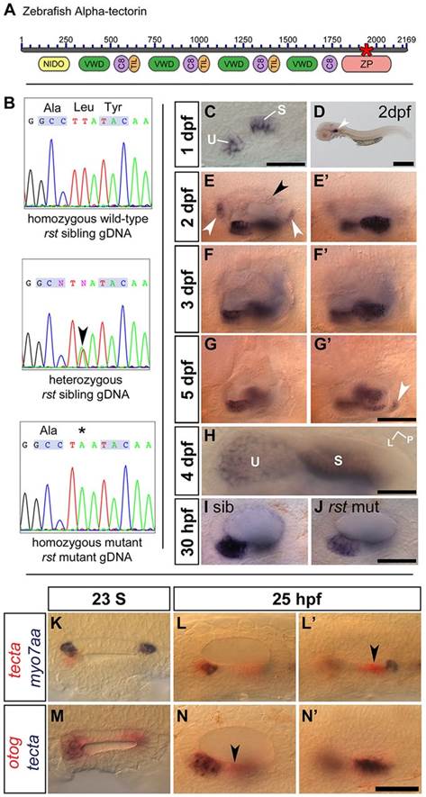
The rst mutation corresponds to a lesion in tecta. (A) Overview of α-Tectorin protein structure based on available sequence data; the N terminus is likely to be incomplete. The asterisk shows the location of the predicted truncation in the rsttl20e allele. NIDO, nidogen domain; VWD, Von Willebrand factor type D domain; C8, domain containing eight conserved cysteine residues; TIL, trypsin inhibitor-like cysteine-rich domain; ZP, zona pellucida domain. (B) Sequencing data from rst mutant, heterozygous sibling and homozygous wild-type sibling embryos showing the T-to-A transversion (arrowhead and asterisk). (C) tecta mRNA expression in the OV of a 1dpf phenotypically wild-type sibling embryo from an eis cross. tecta is expressed in the utricular (U) and saccular (S) maculae. (D) 2dpf wild-type (AB strain) embryo showing that tecta is expressed exclusively in the OV (arrowhead). (E,E2) Within the OV at 2dpf, tecta is expressed at high levels in the utricle and saccule, with lower levels in the semicircular canal projections (black arrowhead) and at the base of the cristae (white arrowheads). (F,F2) Strong tecta expression continues in the utricular and saccular maculae at 3dpf. (G,G2) The level of tecta expression in the maculae is reduced at 5dpf, and a new region of expression appears (arrowhead). (H) Dorsal view of utricle (U) and saccule (S) in 4dpf wild-type embryo. L, lateral; P, posterior. Expression in the utricular macula is strongest at the periphery. (I,J) rst mutants show a lower level of tecta expression than siblings at 30hpf. Genotypes were confirmed by sequencing gDNA. (K) Dorsal view of a 23S wild-type OV, showing that tecta mRNA expression (red) is not restricted to the tether cells (myo7aa mRNA, blue) at seeding stages. (L,L′) Lateral views of 25hpf phenotypically wild-type OV. The focal plane shows the region of the anterior macula in L and the region of the posterior macula in L′. tecta expression includes the tether cells and surrounding cells of the anterior macula (L) and a region just anterior to the tether cells of the posterior macula (arrowhead, L′). (M) Dorsal view of a 23S wild-type OV, showing that otog (red) and tecta (blue) expression roughly overlap at this stage. (N,N′) Lateral views of a 25hpf phenotypically wild-type OV. The focal plane shows the region of the anterior macula in N and the region of the posterior macula in N′. otog (red) is expressed in a tecta-negative area of the ventral OV epithelium (arrowhead, N). Scale bar: 50µm in C,I,J,K-N&prime& 500µm in D; 33µm in H; 75µm in E-G′.