
Fig. 5
|
Fig. 5
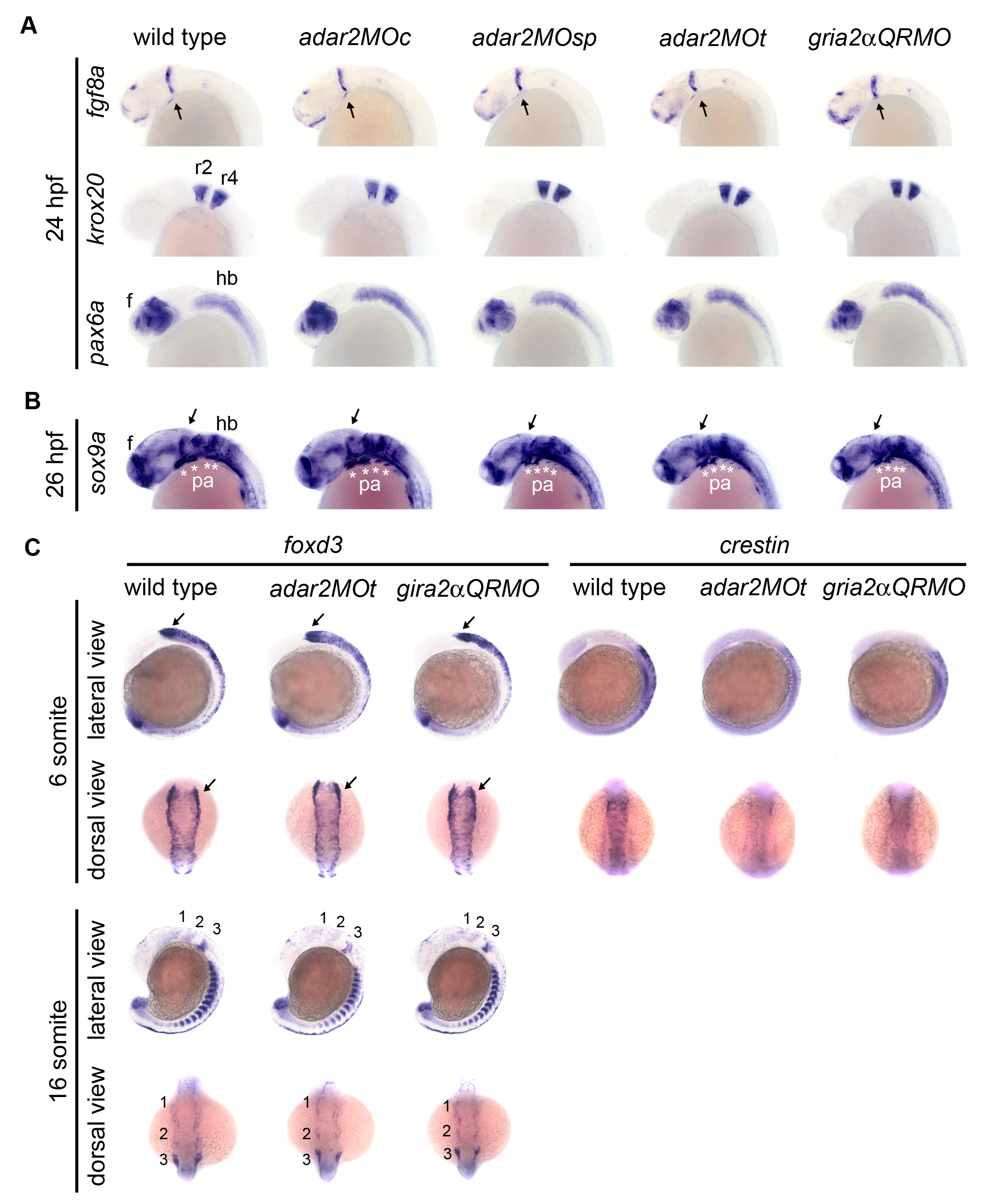
Gene expression in neural tube and migratory neural crest.
(A, B) Embryos are under lateral view. (A) Expressions of brain regionalization genes. Expression of fgf8, krox20, and pax6a appear normal in the 24 hpf hypo-Q/R-editing morphants. (B, C) Expressions of neural crest genes. The expression of mesenchymal condensations marker, sox9a, in the pharyngeal arch (pa, *) are slightly but consistently reduced in the hypo-Q/R-editing morphants. Expressions of neural crest markers foxd3 and crestin are mildly affected at 6-somite and 16-somite stages. Anterior is respectively to the left and top at lateral and dorsal views. 1, 2 and 3 are the three migration cranial neural crest streams originated from posterior mesencephalon and hindbrain. Arrows indicate the midbrain hindbrain boundary. e, eye; hb, f, forebrain, hb, hindbrain; r2 and r4, rhombomeres 2 and 4.