
Fig. 2
|
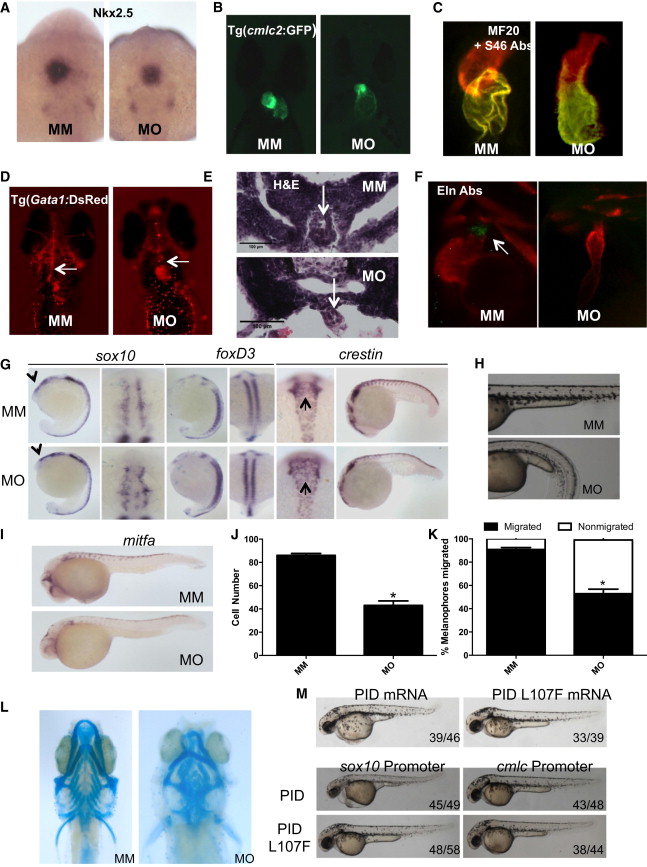
Fig. 2
Pak1 Morphants Display Cardiac and Neural Crest Defects
(A) Transcript expression of nkx2.5 shown by in situ hybridization at 20 somites.
(B and C) Pak1 morphant heart does not loop at 48 hpf as shown by (B) Tg(cmlc2:GFP) immunofluorescence and (C) staining with MF20 and S46 antibodies.
(D) Ventral images of 48 hpf Tg(Gata1:DsRed) show a cardiac outflow tract blockage with no blood flow throughout the head vasculature in the pak1 morphants (MO) compared to MM embryos (MM). Arrows point to cardiac outflow tract.
(E) Transverse sections of the cardiac outflow tract in MO- and MM-injected embryos stained with H&E. Arrows point to the cardiac outflow tract.
(F) Eln2 staining of WT and pak1 morphant embryos. Arrow indicates region of Eln positivity. Eln Abs, Eln2 antibodies.
(G) In situ hybridization for sox10 and foxD3 at 16–18 somites (lateral and cranial views). Arrows point to cranial neural crest expression in lateral view. In situ hybridization for crestin at the 16- to 18-somite stage and the 20-somite stage. Note lack of migration of neural crest in cranial view (arrows) and down the tail in the lateral view.
(H) Trunk images of pak1 morphants lacking melanophores at 2 dpf.
(I) In situ hybridization for mitfa.
(J) Quantification of melanophores at 2 dpf. Error bars indicate a significant difference with p < 0.0001. p < 0.0001.
(K) Quantification of melanophore migration in pak1 morphants compared to MM-injected embryos. Error bars indicate a significant difference with p < 0.0001.
(L) Cartilage staining (Alcian blue) at 6 dpf.
(M) Top: WT embryos were injected with either PID (Pak inhibitor) or PID L107F (inactive Pak inhibitor) mRNA. Embryos are shown at 48 hpf. Bottom: WT embryos were injected with expression vectors encoding PID or PID L107F driven by the sox10 promoter or the cmlc promoter, as indicated. Images were taken at 48 hpf.
Reprinted from Developmental Cell, 29, Kelly, M.L., Astsaturov, A., Rhodes, J., Chernoff, J., A Pak1/Erk Signaling Module Acts through Gata6 to Regulate Cardiovascular Development in Zebrafish, 350-9, Copyright (2014) with permission from Elsevier. Full text @ Dev. Cell