
Fig. S1
|
Fig. S1
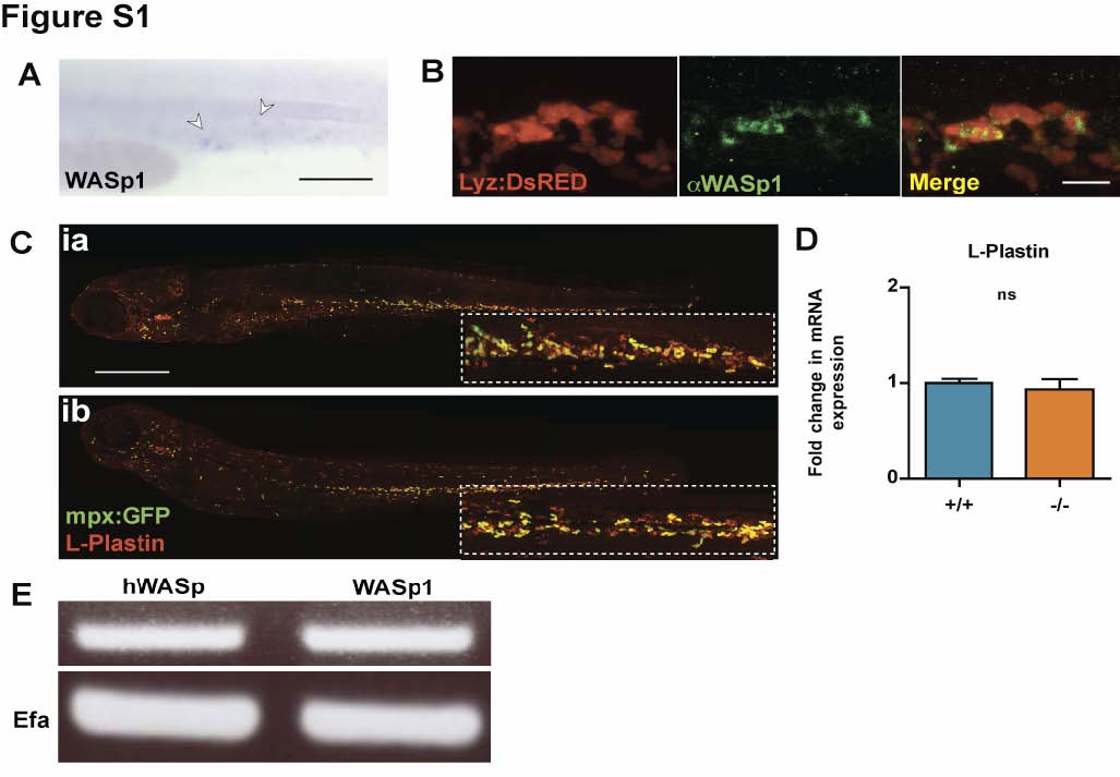
WASp1 is expressed in the hematopoietic lineages of Zebrafish larvae with WASp mutants showing no developmental defect in leukocyte numbers or distribution. (A) Whole-mount in situ hybridization for WASp1 in 1 dpf larvae, indicating expression in the early hematopoietic lineage (white arrowheads). (B) WASp1 antibody immunostaining depicting co-localisation with the transgenic neutrophil marker, lyz:DsRED in 3 dpf larvae. (C) WASp1 mutant has a normal distribution of leukocytes, as shown by L-Plastin immunostaining (red) in an mpx:GFP neutrophil transgenic line (green). (D) WASp1 mutant has a normal level of expression of the leukocyte marker Lplastin at 3 dpf, as shown by qPCR, indicating normal numbers of leukocytes. (E) RT-PCR showing transgenic expression of hWASp in “rescued” larvae is comparable to native levels of WASp1 transcript in WT larvae, with corresponding EFα control. Error bars (s.d). Scale bars: A, 100 μm; B, 10 μm; C, 200 μm.