
Fig. 7
|
Fig. 7
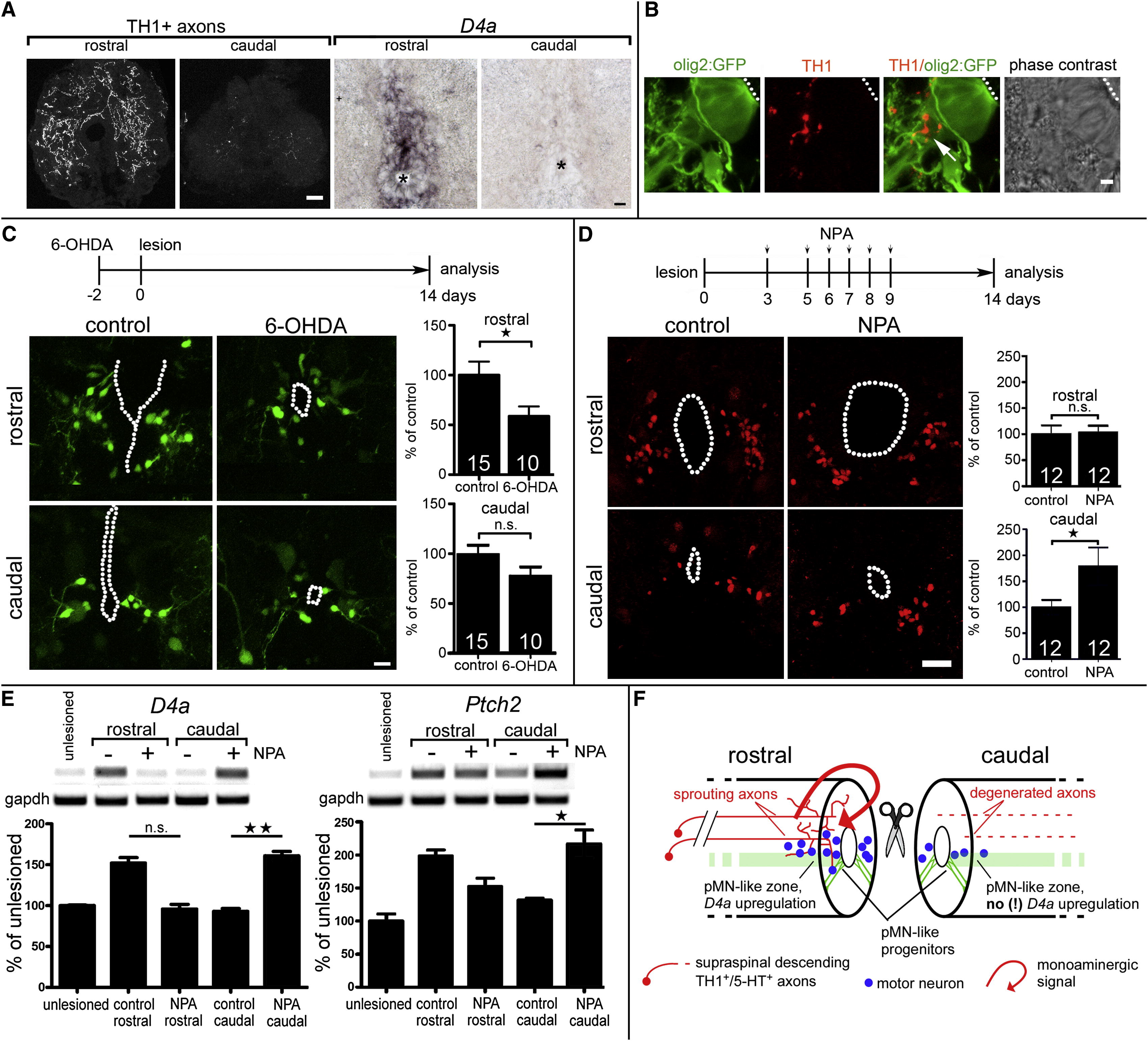
Dopamine Promotes Motor Neuron Regeneration in the Adult Spinal Cord Spinal cross-sections are shown at 14 dpl; central canal is indicated by dots or asterisks. (A) TH1+ axons and d4a mRNA expression are detectable mainly rostral to a spinal lesion site. (B) Higher magnification shows apposition (arrow) of TH1+ axons with radial processes of olig2:GFP+ motor neuron progenitor cells. (C) Ablation of dopaminergic axons with 6-OHDA reduces (p < 0.05) numbers of newly generated HB9:GFP+ motor neurons only rostral to a spinal lesion site. (D) NPA injections significantly (p < 0.05, one sided) increase the number of HB9+ motor neurons only caudal to a spinal lesion site. (E) NPA injections increase d4a (left) and patched2 (right) expression only caudal to the lesion site. Example gels and quantitative densitometric analyses are shown (p < 0.05, p < 0.01; Krukall-Wallis with Dunn’s posttest). (F) Schematic summary of dopamine signaling in relation to motor neuron regeneration at 14 dpl. Error bars represent SEM. Scale bars, 40 μm (A, left), 20 μm (A, right), 5 μm (B), and 15 µm (C and D). See also Figure S6.
Reprinted from Developmental Cell, 25(5), Reimer, M.M., Norris, A., Ohnmacht, J., Patani, R., Zhong, Z., Dias, T.B., Kuscha, V., Scott, A.L., Chen, Y.C., Rozov, S., Frazer, S.L., Wyatt, C., Higashijima, S., Patton, E.E., Panula, P., Chandran, S., Becker, T., and Becker, C.G., Dopamine from the Brain Promotes Spinal Motor Neuron Generation during Development and Adult Regeneration, 478-491, Copyright (2013) with permission from Elsevier. Full text @ Dev. Cell