
Fig. 5
|
Fig. 5
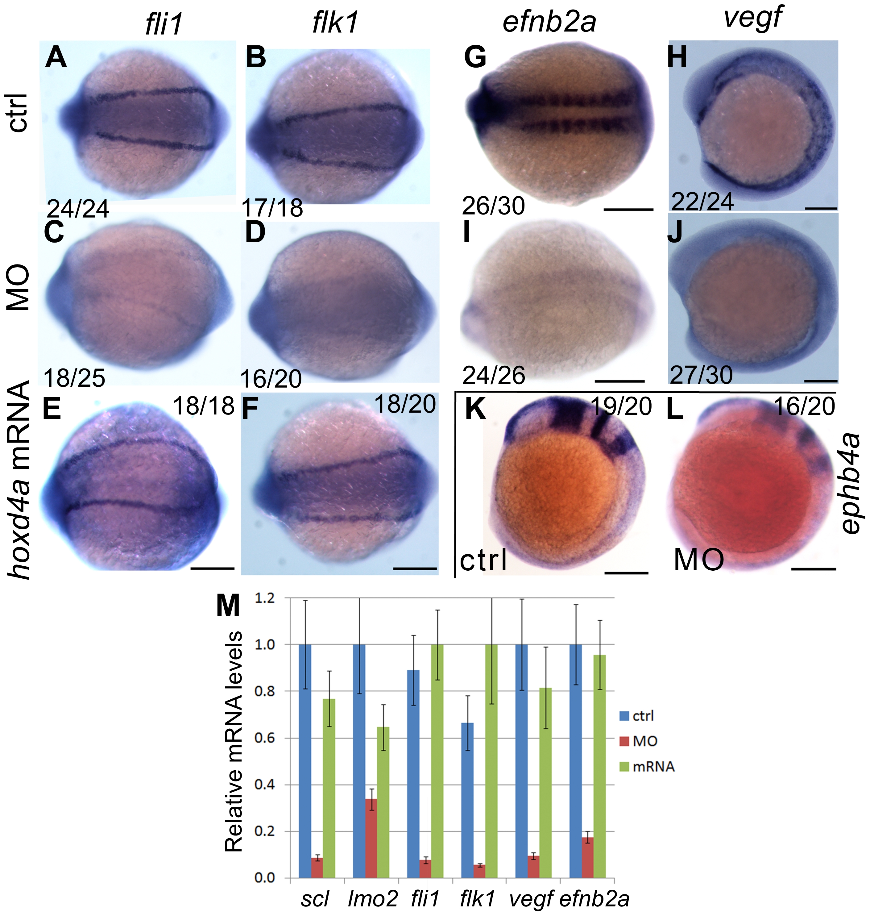
Knockdown of hoxd4a disrupts the endothelial programme in zebrafish embryos.
In situ hybridization at 13 hpf revealing expression of pan-endothelial markers fli1 and flk1 in control-injected embryos (A,B), hoxd4a morphants (C,D), and rescued embryos co-injected with anti-hoxd4a MO and capped mRNA for hoxd4a (E,F). (G,I) Expression of the marker of arterial identity efnb2a in controls (G) and hoxd4a morphants (I). (H,J) Expression of the endothelial inducer vegf in controls (H) and hoxd4a morphants (J). (K,L) Expression of the venous marker ephb4a in controls (K) and hoxd4a morphants (L). Ratios indicate the fraction of embryos showing the presented phenotype. All images show dorsal views with anterior to the left except H, J, K and L which are lateral views with anterior to the left. Scale bars equal 100 μm. (M) qRT-PCR results showing depletion at 26–28 hpf of angioblast and vascular gene expression in hoxd4a morphants and rescue by co-injection of capped mRNA for hoxd4a. Samples were normalized to β-actin. Error bars indicate standard error. By comparison to controls and rescuants, the gene expression levels of all morphants were statistically different to pd0.02 except for lmo2 control vs morphant (p = 0.04) and lmo2 rescuant vs morphant (p = 0.05).