
|
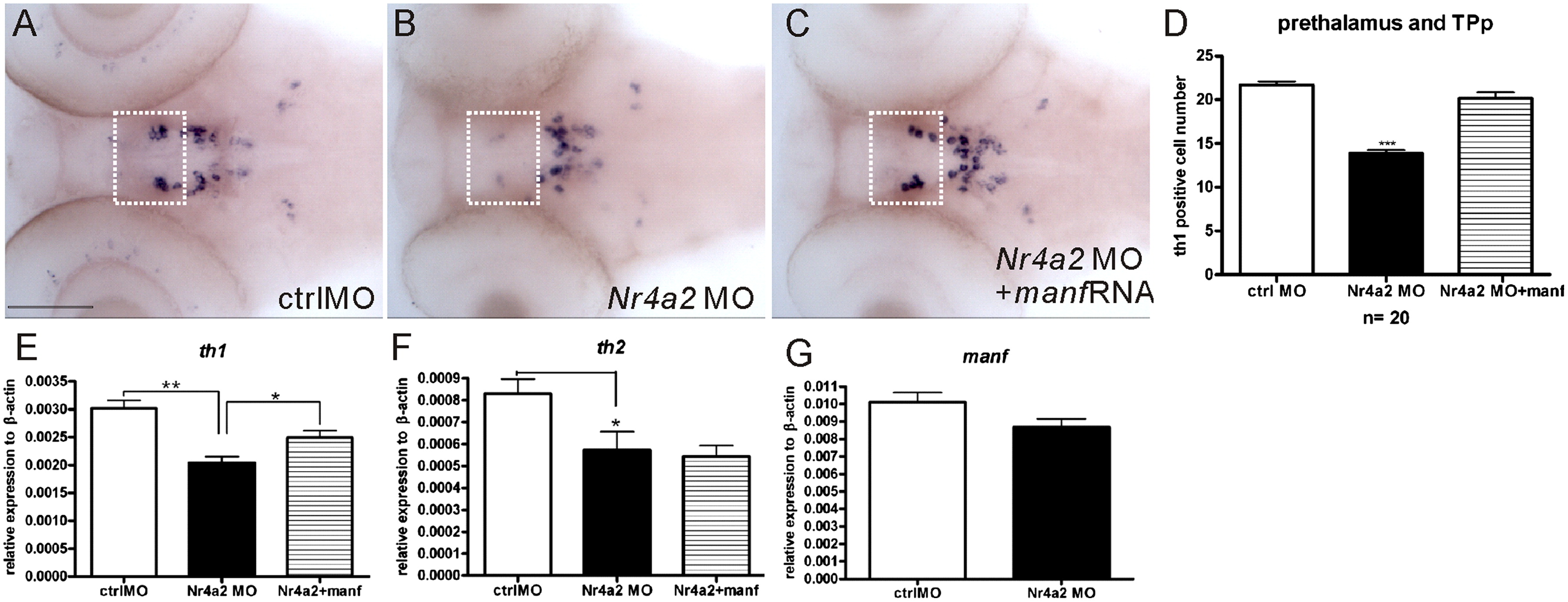
Fig. 8 MANF partially rescued the loss of th1 cells in 3 dpf Nr4a2 morphants. (A–C) th1 expression pattern was revealed by whole mount in situ hybridization at 3 dpf control MO, Nr4a2 MO and Nr4a2 MO co-injected with manf mRNA embryos, anterior to the left and dorsal to the top. (D) Quantitative analysis of the number of th1-containing cells in prethalanus and TPp dopaminergic population 5,6,11 and 12 defined by Sallinen et al., 2009b; DC1 and DC2 group defined by Rink and Wullimann, 2002. (***p<0.001, one-way ANOVA with Dunnett′s test) (E and F) qPCR analysis of th1, th2 and manf transcript levels (*p<0.05, n=3, Student′s t test). White rectangles indicate the loss of th1 cell population in population 5,6,11 and 12. TPp: periventricular nucleus of posterior tuberculum. Scale bar=100 μm.
Reprinted from Developmental Biology, 370(2), Chen, Y.C., Sundvik, M., Rozov, S., Priyadarshini, M., and Panula, P., MANF regulates dopaminergic neuron development in larval zebrafish, 237-249, Copyright (2012) with permission from Elsevier. Full text @ Dev. Biol.