
Fig. 7
|
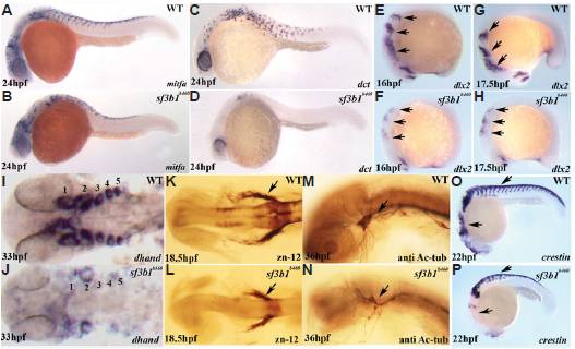
Fig. 7 Neural crest progenitor and cranial ganglia phenotypes of sf3b1b460 mutants. There is reduced melanoblast expression of mitfa (A,B) and nearly absent melanoblast expression of dct (C,D) in sf3b1b460 mutants. (E-J) Pharyngeal arches are dramatically reduced in sf3b1b460 mutant embryos. (E-H) Lateral view of whole-mount in situ embryos with dlx2 antisense RNA probe, arch progenitors denoted by arrows. (I,J) Dorsal view of flat-mounted in situ embryos with dhand antisense RNA probe, pharyngeal arches 1 and 2 are decreased and 3, 4 and 5 are absent in sf3b1b460 mutants. (K-N) the cranial ganglia are also affected in sf3b1b460 mutants. (K,L) Whole-mount antibody staining with zn-12. Arrows in (K,L) show the trigeminal ganglia. (M,N) Whole-mount antibody staining with anti-acetylated tubulin antibody. Arrows in (M,N) show the decreased and disorganized trigeminal ganglia in sf3b1b460 mutants compared to wildtype siblings. (O,P) Crestin expressing cells are present in sf3b1b460 mutant embryos. However, these cells are reduced in numbers, particularly in the cranial region and fail to migrate normally in the sf3b1b460 mutant (P) compared to wildtype embryos (O).