
|
Fig. 4
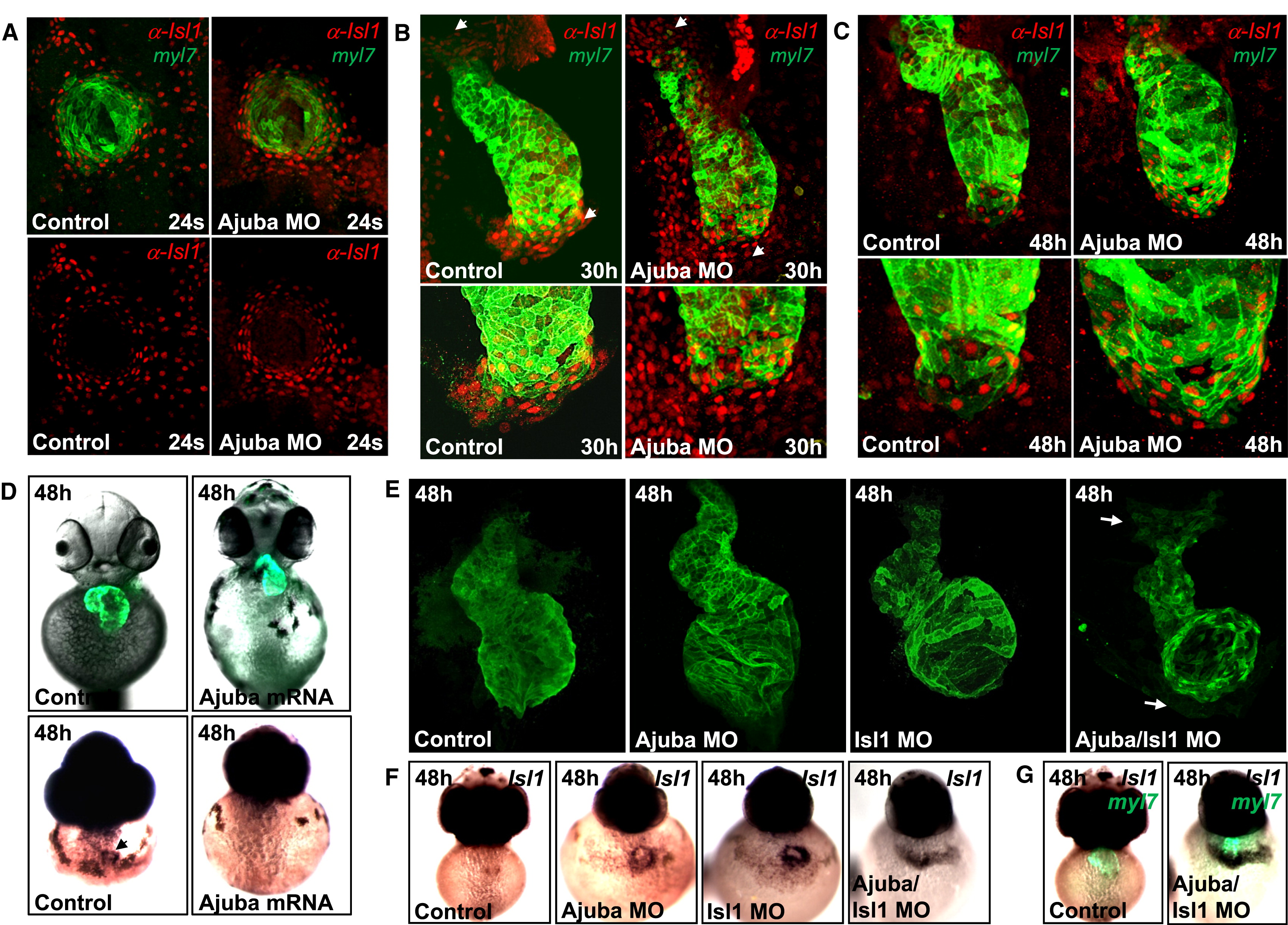
Ajuba and Isl1 Restrict the Number of Isl1-Expressing Cells in the Heart (A) Confocal images of control and Ajuba morphant Tg(myl7:EGFP-HsHRAS)s883 embryos stained with anti-GFP and anti-Isl1 antibodies at 24 somites. (B and C) Confocal images of control and Ajuba morphant Tg(myl7:EGFP-HsHRAS)s883 embryos stained with anti-Isl1 antibodies (red) and visualized by GFP fluorescence (green) for myl7 expression at 30 hpf (B) and at 48 hpf (C). (D) Control and zAjuba mRNA-injected Tg(myl7:EGFP-HsHRAS)s883 embryos at 48 hpf, frontal view (top); in situ hybridization for Isl1 in control or zAjuba mRNA-overexpressing embryos at 48 hpf (bottom). (E) Confocal images of hearts of control, Ajuba, Isl1, and Ajuba/Isl1 morphant Tg(myl7:EGFP-HsHRAS)s883 embryos, stained with anti-GFP antibody. Arrows show differentiated cardiomyocytes in the pericardial wall adjacent to the heart. (F) In situ hybridization for Isl1 in control, Ajuba, Isl1, and Ajuba/Isl1-deficient embryos. (G) Immunostaining with anti-GFP and in situ hybridization for Isl1 in control (left) and Ajuba/Isl1-deficient Tg(myl7:EGFP-HsHRAS)s883 (right) embryos from (F). See also Figure S3.
Reprinted from Developmental Cell, 23(1), Witzel, H.R., Jungblut, B., Choe, C.P., Crump, J.G., Braun, T., and Dobreva, G., The LIM Protein Ajuba Restricts the Second Heart Field Progenitor Pool by Regulating Isl1 Activity, 58-70, Copyright (2012) with permission from Elsevier. Full text @ Dev. Cell