
Image
|
Figure Caption
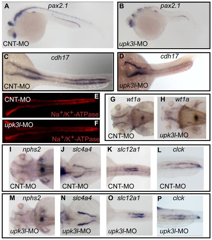
Fig. 4 Pronephric morphogenesis, patterning, and segmentation in control and morphant embryos.
(A–D) Expression of pax2.1 in 1-dpf embryos or cdh17 in 2-dpf embryos as revealed by in situ hybridization. (E, F) Low magnification overview of Na+/K+-ATPase expression in the PTs of 2-dpf control or morphant embryos. (G–P) Expression of wt1a (G–H), nph2s (I, M), slc4a4 (J,N), slc12a (K, O) or clck (L, P) in control or morphant 3-dpf embryos.
Figure Data
Acknowledgments
This image is the copyrighted work of the attributed author or publisher, and
ZFIN has permission only to display this image to its users.
Additional permissions should be obtained from the applicable author or publisher of the image.
Full text @ PLoS One