
|
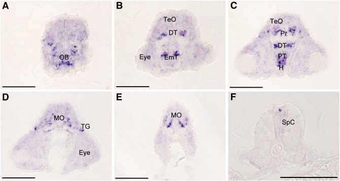
Fig. 4 Expression of 5-HT2C receptor as detected in sectioned whole-mount in-situ hybridization preparations. A set of sections (10 μm) in an anterior to posterior sequence (A to F) from the same preparation is shown. Strong labeling of cell bodies is found in the olfactory bulb (OB in A), the dorsal thalamus (DT) and eminentia thalami (EmT) in a more posterior section (B). The strong staining of neurons in the pretectum (Pr), posterior tuberculum (PT) and hypothalamic area (H) is seen in C. A central cluster of cells stretches from the anterior to the posterior region of the medulla oblongata (MO in D and E). In the posterior region of the medulla, the cells are located in the ventral region and are organized in dorso-ventral stripes (E). (F) The spinal cord (SpC) shows labeling only in isolated pairs of cells at the dorsal tip of the spinal cord. TG — trigeminal ganglion. Scale bar 100 μm (A-E), 50 μm (F).
Reprinted from Gene, 502(2), Schneider, H., Fritzky, L., Williams, J., Heumann, C., Yochum, M., Pattar, K., Noppert, G., Mock, V., and Hawley, E., Cloning and expression of a zebrafish 5-HT(2C) receptor gene, 108-117, Copyright (2012) with permission from Elsevier. Full text @ Gene