
|
Fig. 6
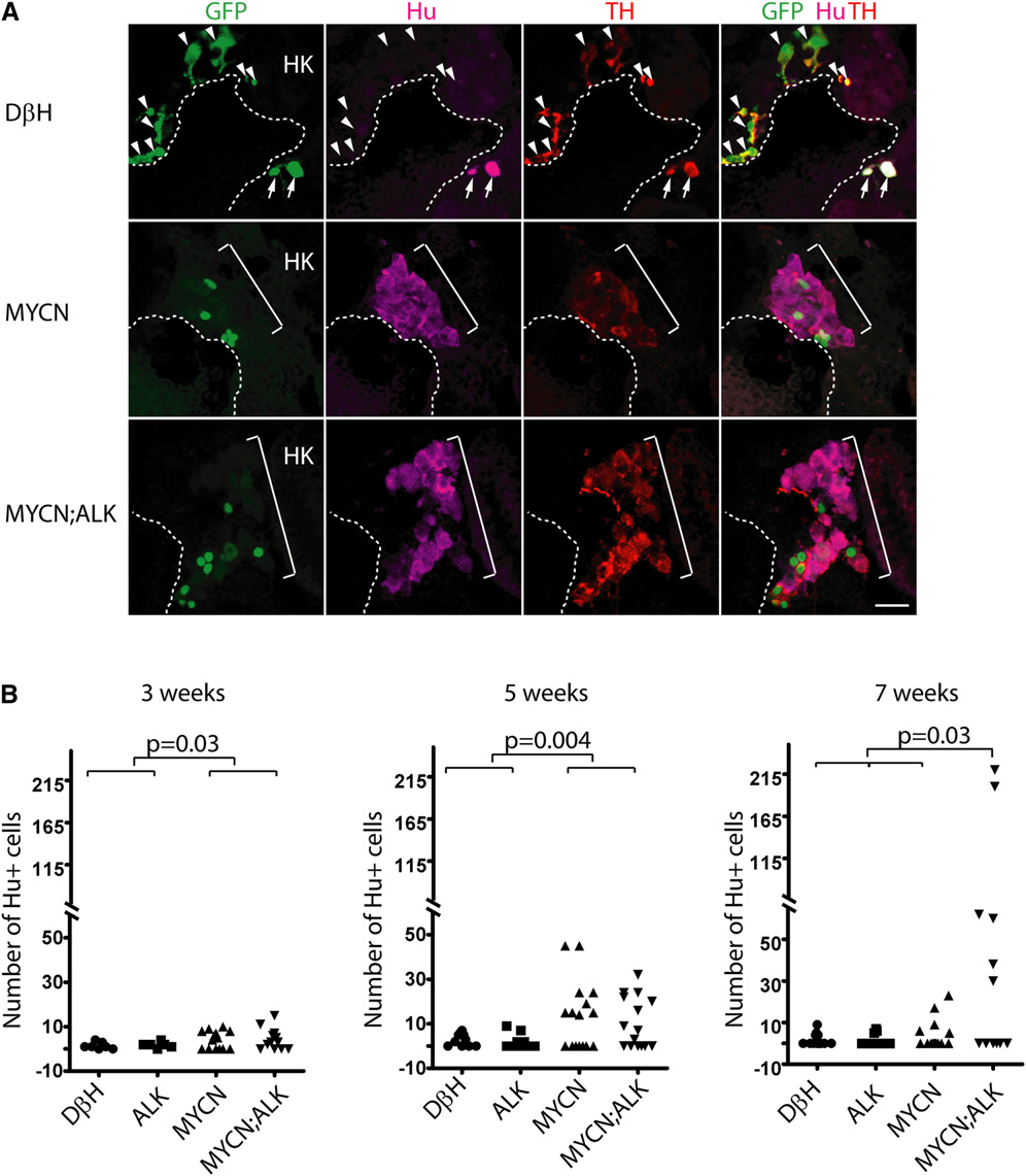
MYCN Causes Hu+ Cell Hyperplasia in the Interrenal Gland (A) Sagittal sections through the interrenal gland in DβH (top panels), MYCN (middle panels), and MYCN;ALK (lower panels) transgenic fish at 5wpf (dorsal up, anterior left). EGFP, green; Hu, magenta; TH, red. Representative sections through the interrenal gland in DβH fish contain three to five GFP+/Hu+/TH+ sympathetic neuroblasts (arrows) and many GFP+/Hu-/TH+ chromaffin cells (arrowheads). Hu+ cell numbers increase in MYCN and MYCN;ALK fish (brackets), and can be GFP+ and TH+. Dotted lines indicate the head kidney (HK) boundary. Scale bar represents 20 μm. (B) Numbers of Hu+ interrenal gland cells in DβH, ALK, MYCN, and MYCN;ALK transgenic fish at 3, 5, and 7 weeks. Means of Hu+ cell numbers were compared by the two-tailed Wilcoxon signed-rank test. See also Figure S6.
Reprinted from Cancer Cell, 21(3), Zhu, S., Lee, J.S., Guo, F., Shin, J., Perez-Atayde, A.R., Kutok, J.L., Rodig, S.J., Neuberg, D.S., Helman, D., Feng, H., Stewart, R.A., Wang, W., George, R.E., Kanki, J.P., and Look, A.T., Activated ALK Collaborates with MYCN in Neuroblastoma Pathogenesis, 362-373, Copyright (2012) with permission from Elsevier. Full text @ Cancer Cell