
|
Fig. 1
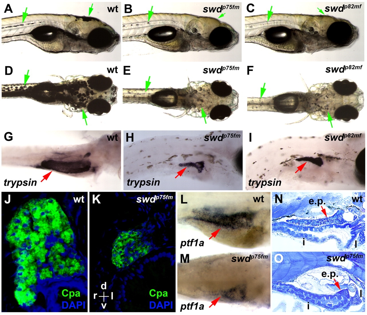
The zebrafish swdp75fm and swdp82mf mutations cause exocrine pancreatic hypoplasia and reduced skin pigmentation. (A–F) Bright-field images of the swd mutants and wild-type (wt) larvae. Green arrows point to pigmented skin. (G–I) Exocrine pancreas (red arrows) revealed by whole-mount in situ hybridization using anti-trypsin riboprobes. (J,K) Histological transverse sections of exocrine pancreas following immunohistochemistry using anti-Cpa antibodies. Staining with DAPI was used to visualize the nuclei. The histological sections are oriented as indicated: d, dorsal; v, ventral; r, right; l, left. (L,M) Exocrine pancreas (red arrows) revealed by whole-mount in situ hybridization using anti-ptf1a riboprobes. (N,O) Sagittal histological sections of larvae. e.p., exocrine pancreas; i, intestine; l, liver. (G,L) wt larvae were grown in E3 medium supplemented with PTU, which inhibits skin pigmentation, in order to facilitate visualization of the exocrine pancreas expressing trypsin or ptf1a. Note that the larvae shown in A–O were analyzed on 5 dpf. (A–C,G–I,L–O) The larvae are positioned in the same orientation and viewed from the right lateral side. (D–F) The views are in the dorsoventral direction.