
|
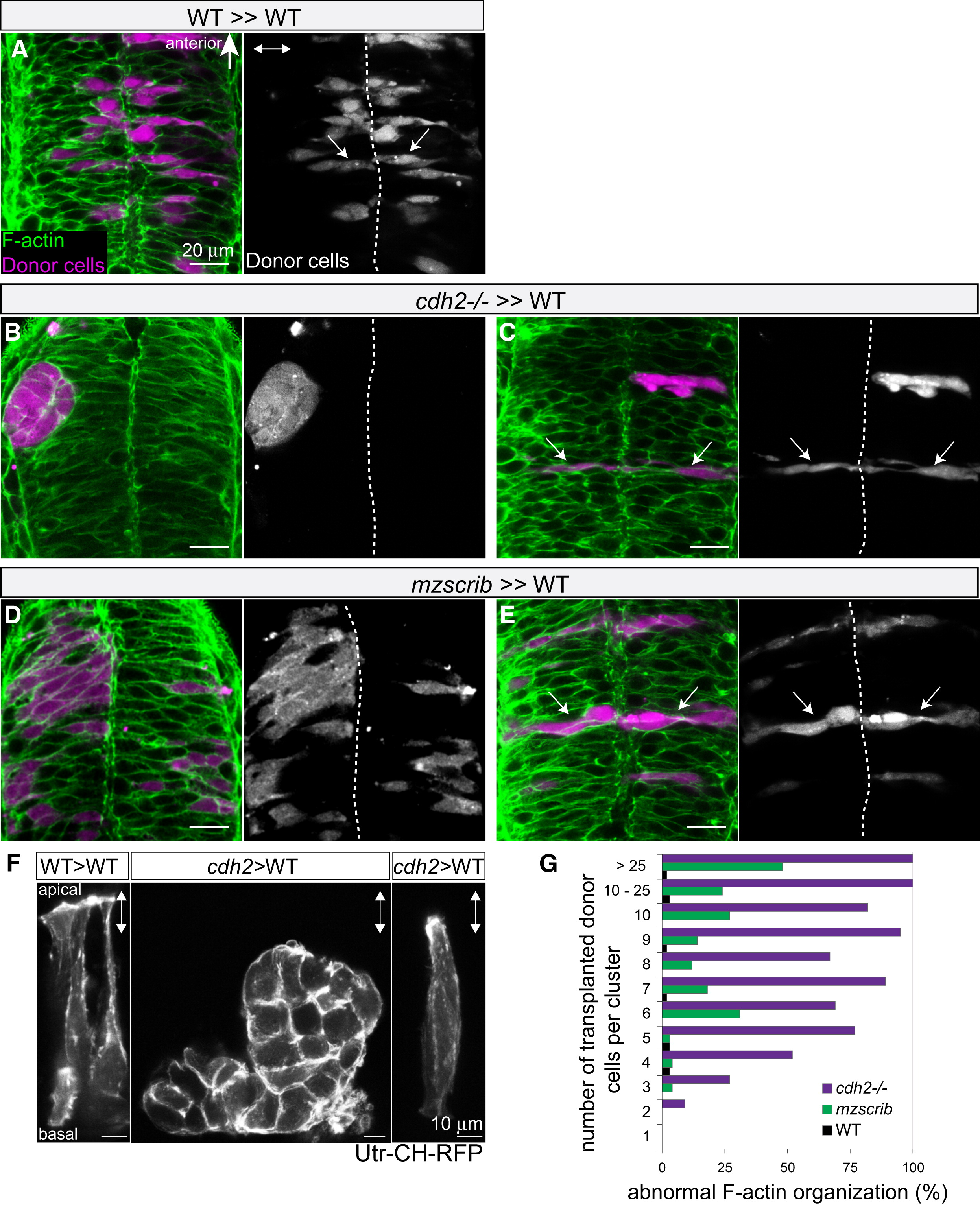
Fig. 4 Nonautonomous Rescue of Oriented Cross-Midline Cell Division of Single cdh2-/- and mzscrib Mutant Cells
(A–E) Genetic mosaics at neural tube stage (21 hpf) in which donor-derived cells (purple) were transplanted into the presumptive posterior hindbrain of WT host embryos (F-actin in green).
(A) Bilateral distribution of WT cells (arrows) in a WT environment.
(B) cdh2-/- cells form unilateral aggregates in a WT environment.
(C) In contrast, isolated cdh2-/- progenitors show rescued bilateral distribution and normal cell shape.
(D) A large group of mzscrib mutant cells in a WT environment with unilateral cell distribution.
(E) Isolated mzscrib cells in a WT environment have rescued shape and bilateral distribution.
(F) Utr-CH-RFP reporter in mosaic embryos showing normal apical enrichment in WT control transplants, loss of apical enrichment in a cluster of transplanted cdh2-/- cells, and rescue in a single cdh2-/- cell that is surrounded by WT host cells. Images represent maximum-intensity projections of optical sections.
(G) Quantification of occurrence of abnormal F-actin organization in WT (black; n = 5 experiments, 13 embryos), mzscrib (blue; n = 3 experiments, 21 embryos), and cdh2-/- (red; n = 2 experiments, 15 embryos) mosaics.
Embryos are shown in dorsal view with anterior to the top. Dotted white line indicates the neural tube midline; two-way arrows indicate the apicobasal axis of the neuroepithelium. See also Figure S4.