
|
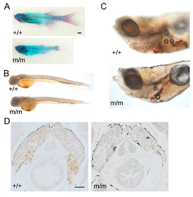
Fig. S1 Phenotypic abnormalities in c-myb mutants. (A) Incomplete ossification in mutant fish (Lower; merged from three pictures) at 9 wk of age as revealed by alcian blue (cartilage) and alizarin red (red) staining. Note the stunted appearance and reduced bone structures. The photograph of the wild-type fish is a composite of two pictures. (Scale bar: 1 mm.) (B) Slightly reduced hemoglobin levels in mutant fish at 2 d postfertilization (dpf) as revealed by whole-mount o-dianisidine staining (orange color). Photographs are composites of three pictures. (C) Reduced hemoglobin levels in mutant fish at 9 dpf as revealed by whole-mount o-dianisidine staining. Photographs are composites of two pictures. (D) Lack of hemoglobin staining in the head kidney of mutants at 7 wk of age revealed by o-dianisidine staining on tissue sections. (Scale bar: 100 μm.)