
|
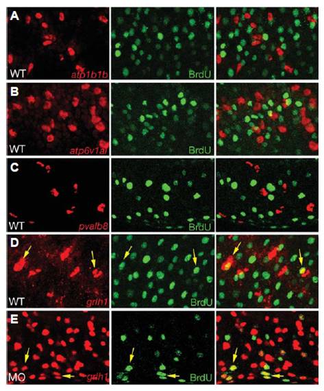
Fig. 5 grhl1 is expressed in proliferating cells. All panels show confocal sections of embryos at 24 hpf that had been incubated with BrdU from 16 - 24 hpf, and after fluorescent in situ hybridization with probes indicated in red (left panels), and after anti-BrdU immunostaining (middle panels); right panels show merged images. (A-D) Wild-type embryos; (E) grhl1 morphant injected with splice-MO (compare with Fig. 6 and Supplementary Fig. S7). Yellow arrows in (D,E) point to grhl1, BrdU-double positive cells. Differentiated ionocytes and pvalb8-positive cells lack BrdU incorporation (A-C), whereas over one quarter of grhl1-positive cells have undergone DNA replication between 16 and 24 hpf (D) (<28%, 17.6±2.83 / 62±4.45, n = 10 sections). (E) In morphant embryos, the absolute number of grhl1-positive cells increases enormously (see also Fig. 6N), whereas the absolute number of grhl1, BrdU-positive cells remains similar (19.3±2.44 / 341±8.91, <5.5%, n = 10 sections), suggesting that the extra grhl1 cells represent differentiated non-keratinocyte epidermal cells, rather than their precursors.