
|
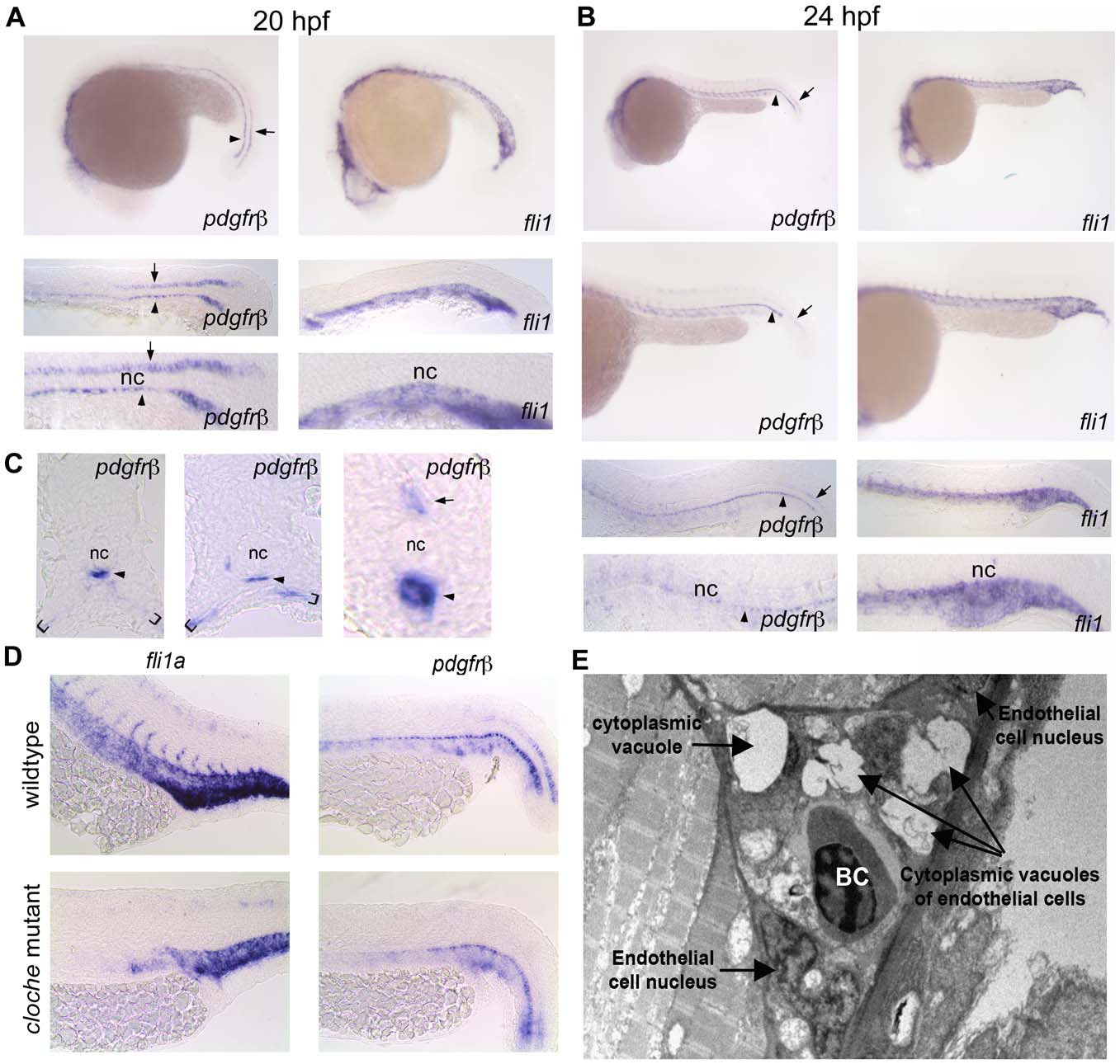
Fig. 2 pdgfrβ2 expression in the developing zebrafish.
In situ hybridization of embryos at 20 hpf (A) and 24 hpf (B) using an anti-sense probe against fli1a or pdgfrβ2. pdgfrβ2 was expressed in the floorplate (arrow) and the hypochord (arrowhead) at 20 hpf. fli1a is known to be expressed in the tail vasculature. Floorplate expression of pdgfrβ2 diminished by 24 hpf while expression in the hypochord persisted and spread ventrally in association with the dorsal aorta and posterior cardinal vein (B). Cross section analysis further indicated pdgfrβ2 expression in the hypochord (arrowhead), floorplate (arrow) and ventral somite boundary (brackets). cloche mutant embryos showed normal expression of pdgfrβ2 indicating that pdgfrβ2 was not expressed in endothelial cells of the dorsal aorta or posterior cardinal vein (D). Transmission electron micrograph images of a longitudinal section through the ISVs (E). Mural cells surrounding the endothelial cells of the ISVs were absent at 72 hpf. Notochord (nc), blood cell (BC).