
|
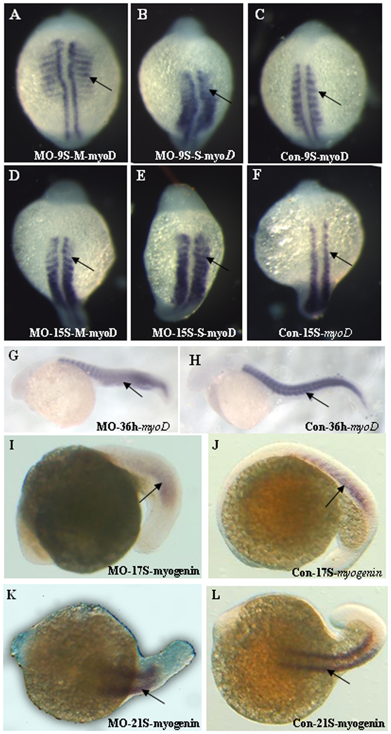
Fig. 11 Skeletal muscle development is altered in Atoh8 knock-down embryos.
(A–H) myoD in situ hybridization; (I–L) myogenin in situ hybridization. (A–C) 9-Somite stage; (D–E) 15-Somite stage; (G–H) 36 hpf; (C,F,H,J and L) are controls and the remainder are morphants with (B) and (E) the severely affected embryos. Expression of the skeletal marker myoD in paraxial mesoderm showed to tendency towards a ventral localization (A,D) or the boundaries of the posterior somites were indistinct(B,C) and a U-shaped somite formation appeared in the morphant (G). Expression of the skeletal marker myogenin provides further evidence of abnormality (I–L).