
IMAGE
Fig. S3
- ID
- ZDB-IMAGE-100520-9
- Publication
- Hong et al., 2010 - The polarity protein Pard3 is required for centrosome positioning during neurulation
- All Figures
- Figures for Hong et al., 2010
Image
|
Figure Caption
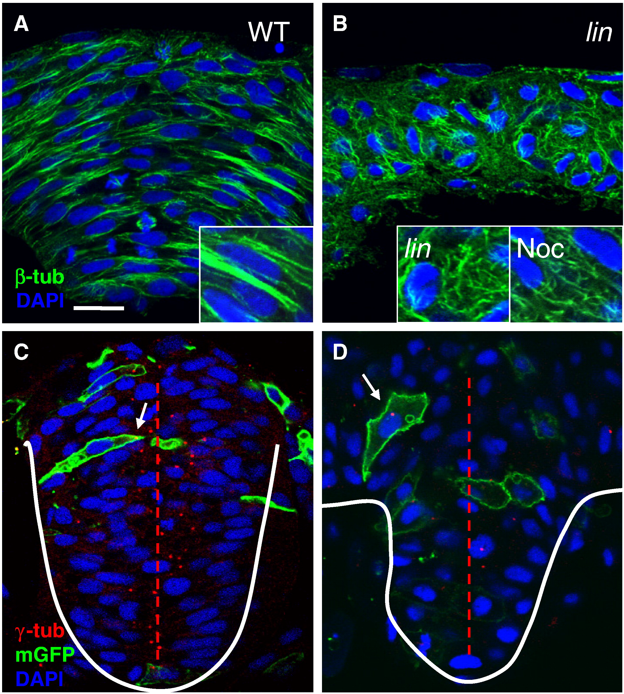
Fig. S3 MT organization and centrosome positioning are abnormal in lin mutants. Cross sections through the hindbrain region of WT (A,C), lin mutant (B, D) and nocodazole (noc)-treated (B, inset) embryos at 4–5 som, immunolabeled with anti-β-tub (A, B) and anti-γ-tub and anti-GFP (C,D). Nuclei are labeled with DAPI. Enlarged views of anti-β-tub labeling. White arrows point toward centrosomes in (C) and (D). Red dotted line depicts the midline. Scale bar: 10 μm.
Figure Data
Acknowledgments
This image is the copyrighted work of the attributed author or publisher, and
ZFIN has permission only to display this image to its users.
Additional permissions should be obtained from the applicable author or publisher of the image.
Reprinted from Developmental Biology, 341(2), Hong, E., Jayachandran, P., and Brewster, R., The polarity protein Pard3 is required for centrosome positioning during neurulation, 335-345, Copyright (2010) with permission from Elsevier. Full text @ Dev. Biol.