
IMAGE
Fig. S2
- ID
- ZDB-IMAGE-100520-8
- Publication
- Hong et al., 2010 - The polarity protein Pard3 is required for centrosome positioning during neurulation
- All Figures
- Figures for Hong et al., 2010
Image
|
Figure Caption
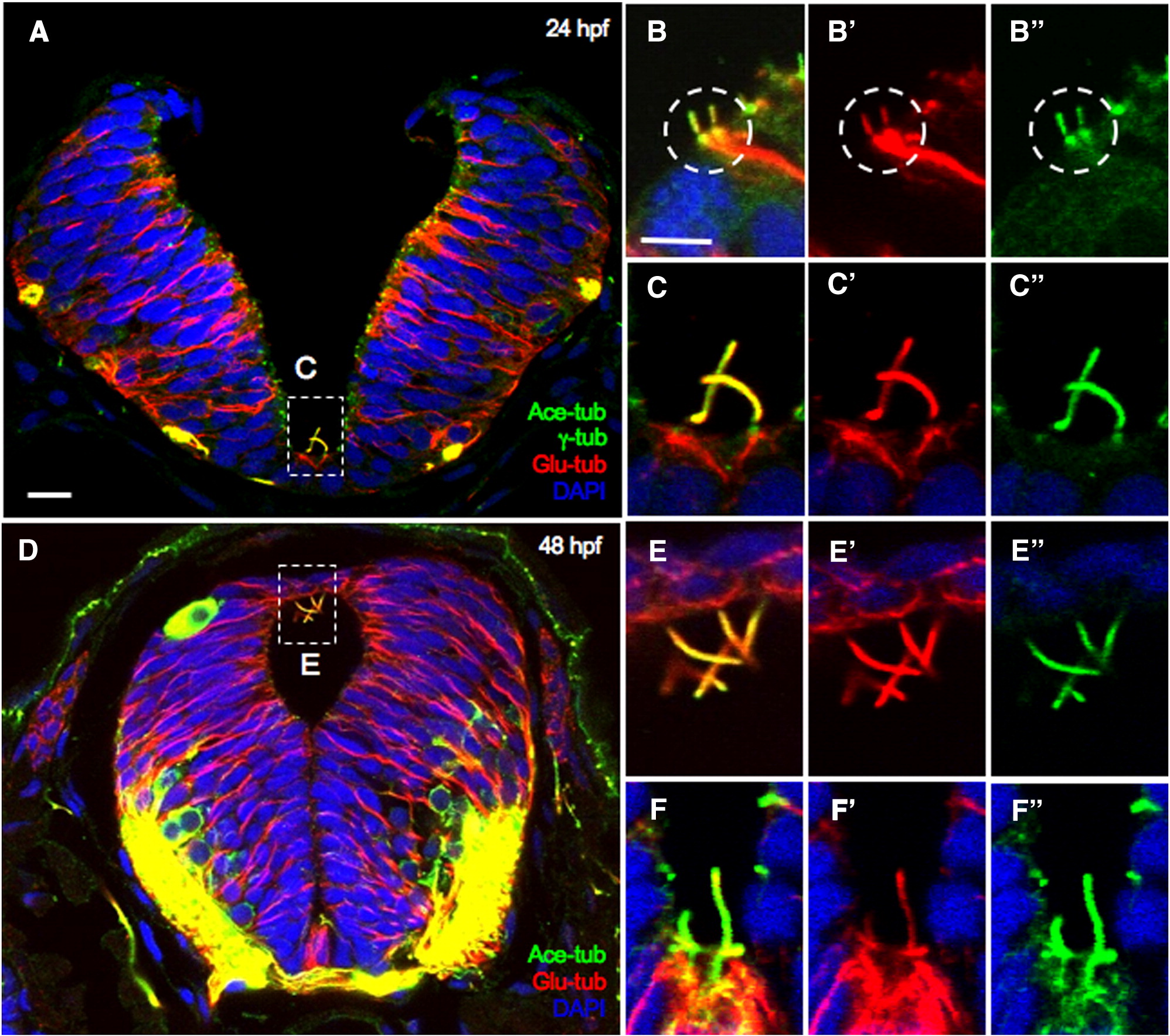
Fig. S2 The centrosome/MT/cilium complex is maintained in the neural tube. Cross Sections through the hindbrain region of 24 hpf (A–C″) and 48 hpf (D–F″) embryos immunolabeled with anti-Glu-tub, anti-Ace-tub and anti-γ-tub (A–C″) or with anti-Glu-tub and anti-Ace-tub (D–F″). Nuclei are labeled with DAPI. Symbols: white boxes in A and D: region that is magnified in (C–C″, E–E″), white circle in B–B″: a centrosome/cilia/MT complex. Scale bars: 10 μm in A and 5 μm in B and E.
Acknowledgments
This image is the copyrighted work of the attributed author or publisher, and
ZFIN has permission only to display this image to its users.
Additional permissions should be obtained from the applicable author or publisher of the image.
Reprinted from Developmental Biology, 341(2), Hong, E., Jayachandran, P., and Brewster, R., The polarity protein Pard3 is required for centrosome positioning during neurulation, 335-345, Copyright (2010) with permission from Elsevier. Full text @ Dev. Biol.