
IMAGE
Fig. 3
- ID
- ZDB-IMAGE-100520-3
- Publication
- Hong et al., 2010 - The polarity protein Pard3 is required for centrosome positioning during neurulation
- All Figures
- Figures for Hong et al., 2010
Image
|
Figure Caption
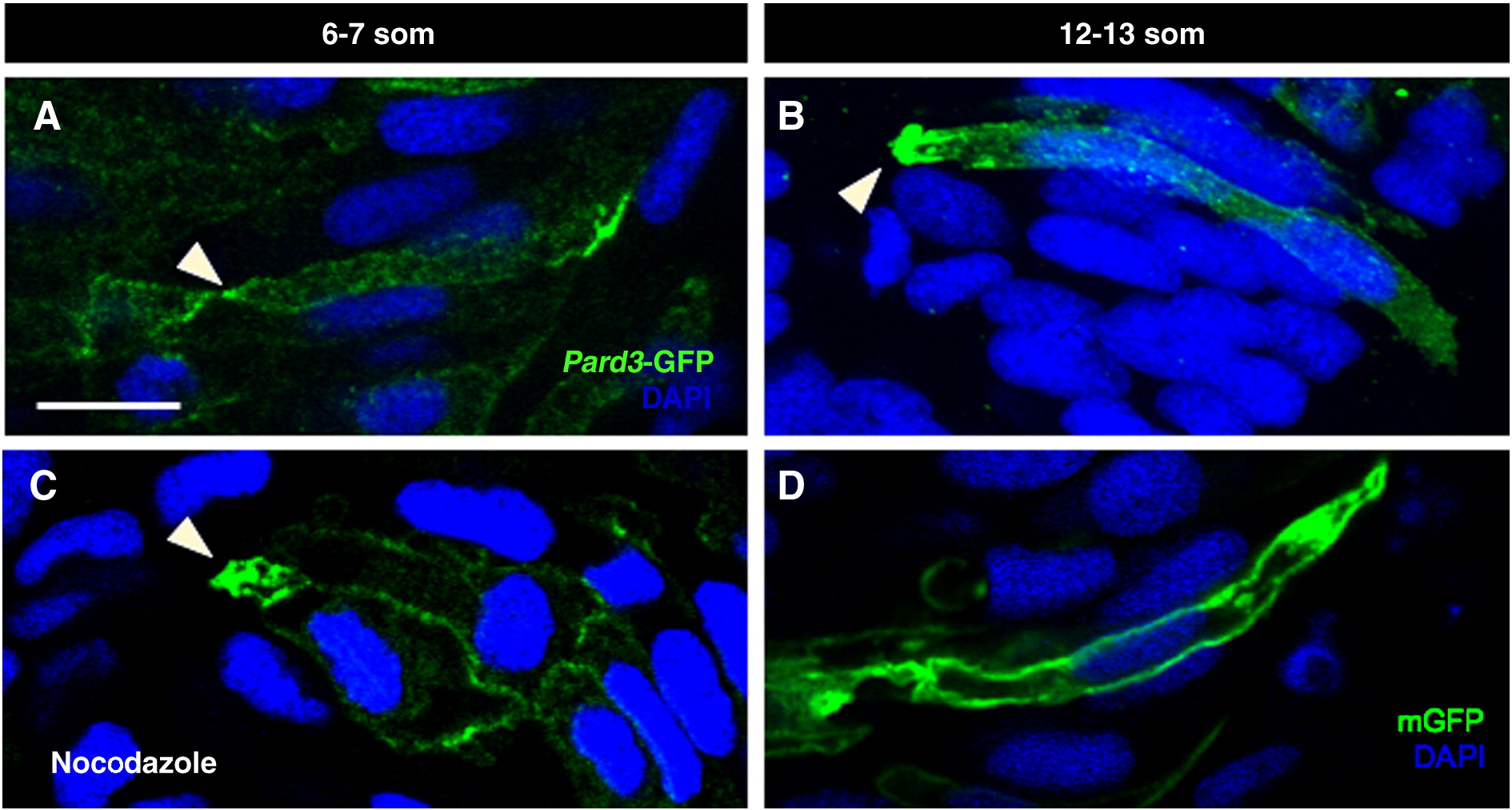
Fig. 3 Localization of Pard3-GFP may be microtubule-independent. Sections of control (A, B, D) and nocodazole-treated (C) embryos at 6–7 som (A, C) and 12–13 som (B, D). (A–C) Embryos were mosaically injected at the 8–16 cell stage with pard3-GFP mRNA and immunolabeled with anti-GFP. (C) The nocodazole treatment was performed for 30 min at an early 6 som stage and the embryo was fixed immediately following treatment. (D) Embryo was injected with mGFP DNA and immunolabeled with anti-GFP. Arrowheads: apical localization of Pard3-GFP. Scale bar: 10 μm.
Acknowledgments
This image is the copyrighted work of the attributed author or publisher, and
ZFIN has permission only to display this image to its users.
Additional permissions should be obtained from the applicable author or publisher of the image.
Reprinted from Developmental Biology, 341(2), Hong, E., Jayachandran, P., and Brewster, R., The polarity protein Pard3 is required for centrosome positioning during neurulation, 335-345, Copyright (2010) with permission from Elsevier. Full text @ Dev. Biol.