
IMAGE
Fig. 2
- ID
- ZDB-IMAGE-100520-2
- Publication
- Hong et al., 2010 - The polarity protein Pard3 is required for centrosome positioning during neurulation
- All Figures
- Figures for Hong et al., 2010
Image
|
Figure Caption
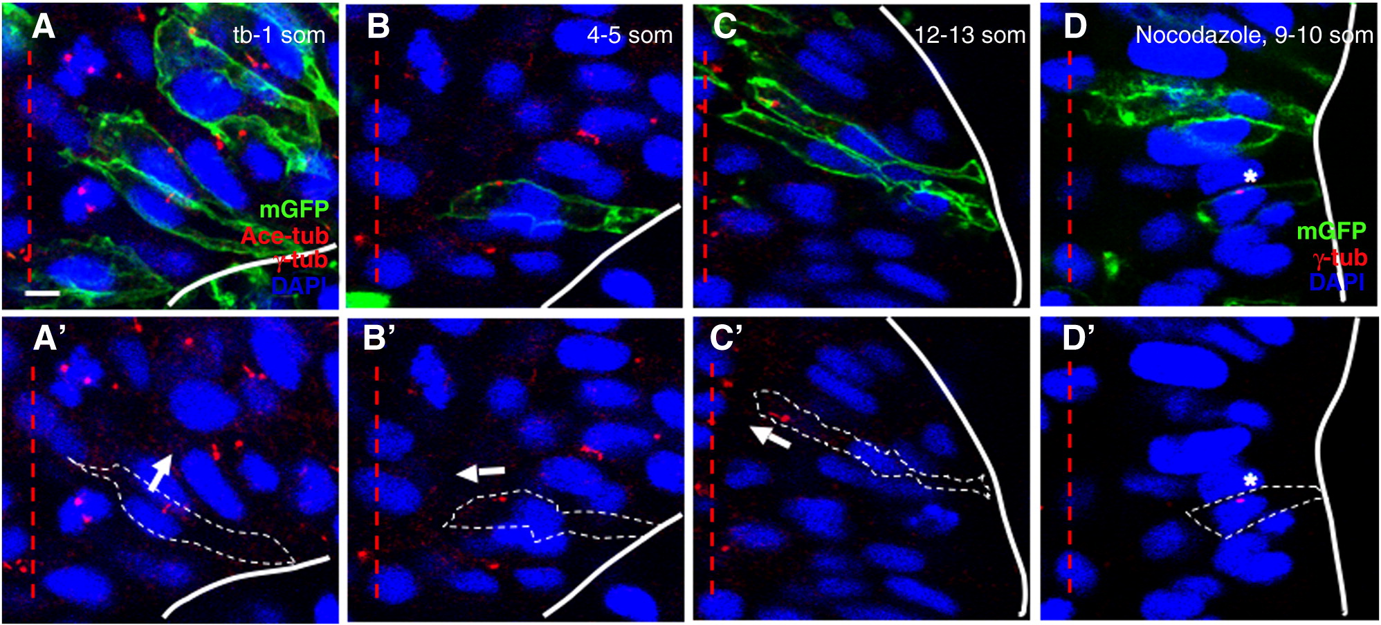
Fig. 2 Centrosome position during neural convergence is microtubule-dependent. Sections of WT (A–C) and nocodazole-treated (D) embryos at the neural plate (A, A′), neural keel (B, B′) and neural rod (C, C′, D, D′) stages immunolabeled with anti-Ace-tub, anti-γ-tub and anti-GFP (A–C) or anti-γ-tub and anti-GFP (D). (A′, B′, C′, D′) Same panels as in (A, B, C, D) without the mGFP label. Red lines: midline; white lines: outline of the cells; arrows: orientation of axonemes; asterisk: ectopic centrosome. Scale bar: 5 μm.
Acknowledgments
This image is the copyrighted work of the attributed author or publisher, and
ZFIN has permission only to display this image to its users.
Additional permissions should be obtained from the applicable author or publisher of the image.
Reprinted from Developmental Biology, 341(2), Hong, E., Jayachandran, P., and Brewster, R., The polarity protein Pard3 is required for centrosome positioning during neurulation, 335-345, Copyright (2010) with permission from Elsevier. Full text @ Dev. Biol.