
Fig. 1
|
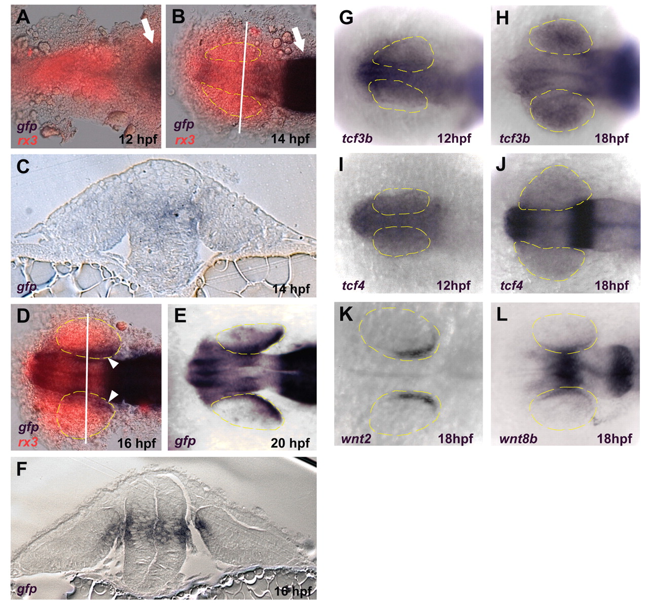
Fig. 1 Wnt signaling becomes active in the dorso-posterior retinal pigmented epithelium (RPE) between 14 and 16 hpf. (A-F) Expression of the TOP:dGFP Wnt reporter detected using in situ hybridization for gfp (blue). In A, B and D, the embryos were also probed for rx3 expression (red) which marks the eye field. (A,B) Dorsal views, anterior left. Active Wnt signaling does not extend rostrally past the midbrain-hindbrain boundary (arrows) at 12 hpf, and approaches but does not enter the eye field at 14 hpf. (C,F) Coronal sections through caudal midbrain/posterior optic vesicles, dorsal up. The lines in B and D indicate the planes of section in C and F, respectively. Active Wnt signaling is seen in the dorsoposterior RPE at 16 hpf, but not at 14 hpf. (D,E) Dorsal views, anterior left. Active Wnt signaling is clearly present in the dorso-posterior eye field at 16 and 20 hpf. (G-L) Dorsal views, anterior left. (G-J) Expression of tcf3b and tcf4 is present in the early eye-field during optic vesicle evagination (12 hpf) and throughout the eye at 18 hpf. (K) The Wnt ligand wnt2 is expressed in the dorsal RPE at 18 hpf. (L) Expression of wnt8b in the midbrain and RPE at 18 hpf.