
|
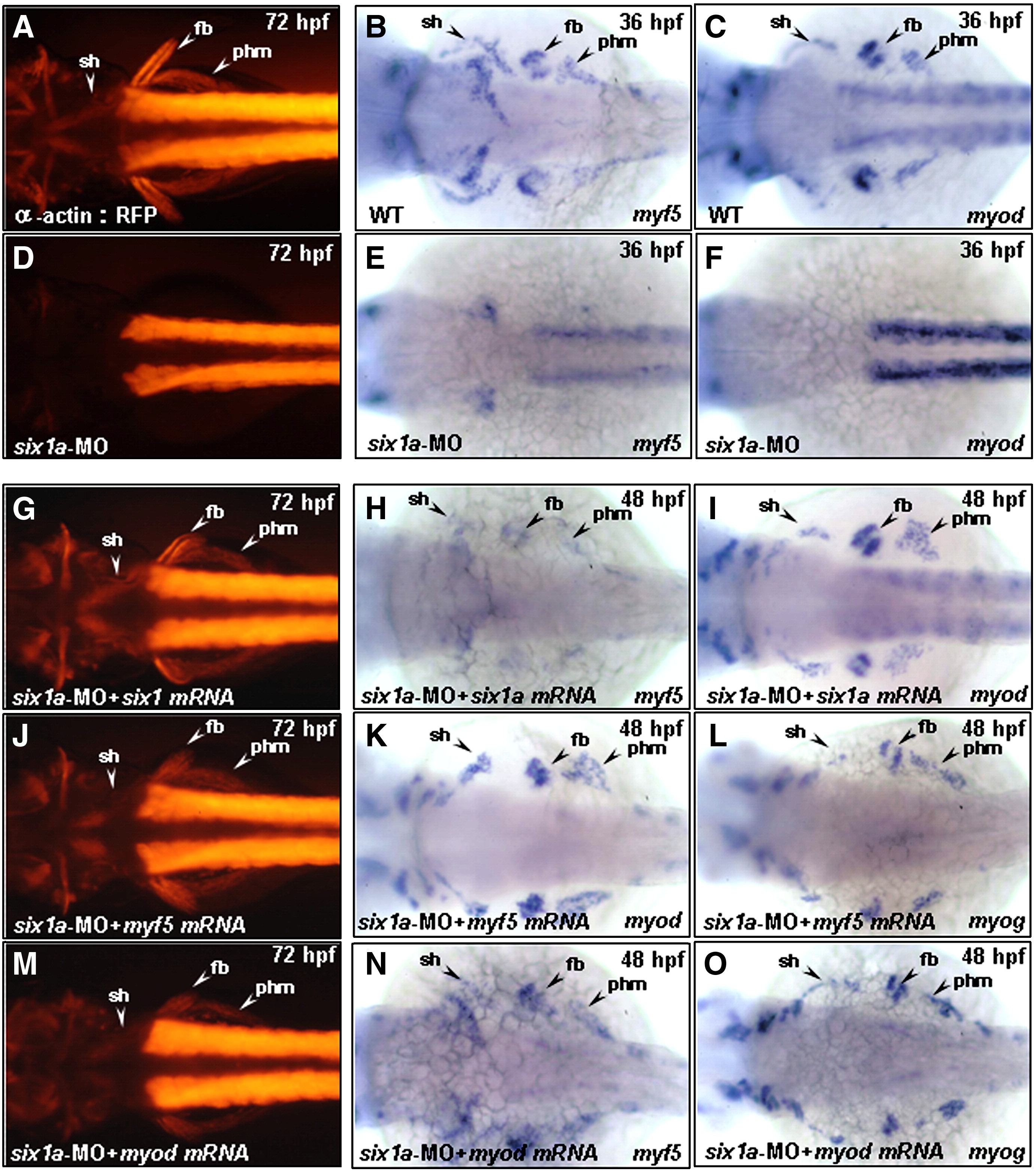
Fig. 5 Injection of six1a-, myf5-and myod mRNA enables embryos to rescue trunk migratory head muscle sternohyoideus (sh) defect in six1a morphants. Dorsal views of embryos derived either from the transgenic line Tg(α-actin:RFP) (A, D, G, J, M) or from the wild-type (B, C, E, F, H, I, K, L). The RFP expression in the embryos derived from the transgenic line at 72 hpf (A) and the detection of myf5 and myod by whole-mount in situ hybridization at 36 hpf (B, C) served as control groups. Injection of embryos with either 8 ng of six1a-MO alone (D–F) or co-injection with 8 ng of six1a-MO and 150 pg of six1a mRNA (G–I), 100 pg of myf5 mRNA (J–L) or 50 pg of myod mRNA (M–O) were examined. RFP, myf5 and myod were not detected in sh, fb, or phm primordia in the six1a morphants (D–F); however, co-injection of six1a mRNA enabled embryos to rescue the defective expressions of RFP, myf5 and myod in sh, fb and phm primordia induced by six1a-MO at 48 hpf (H, I) and at 72 hpf (G). Meanwhile, injection of myf5 mRNA enabled embryos to rescue the defective expressions of RFP, myod and myogenin in sh, fb and phm primordia induced by six1a-MO at 48 hpf (K, L) and at 72 hpf (J). Injection of myod mRNA enabled embryos to rescue the defective expressions of RFP, myf5 and myogenin in sh, fb and phm primordia induced by six1a-MO at 48 hpf (N, O) and at 72 hpf (M). fb, fin bud; phm, posterior hypoaxial muscle; and sh, sternohyoideus.
Reprinted from Developmental Biology, 331(2), Lin, C.Y., Chen, W.T., Lee, H.C., Yang, P.H., Yang, H.J., and Tsai, H.J., The transcription factor six1a plays an essential role in the craniofacial myogenesis of zebrafish, 152-166, Copyright (2009) with permission from Elsevier. Full text @ Dev. Biol.