
|
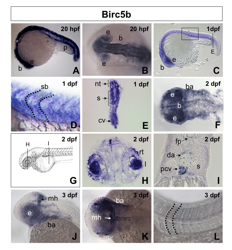
Fig. 1 Birc5b spatiotemporal expression in zebrafish embryos. A, B. Lateral (A) and dorsal (B) views of 20 hpf (20 somites) embryos revealing expression of Birc5b in the neural tube, brain, pronephric duct and eyes. C. Lateral view 1 dpf (30 somites) embryo, showing Birc5b expression in brain, eye, neural tube, somite and intersomite boundaries, with higher magnification in D. E. Transverse section through 1 dpf embryo (from C), revealing expression of Birc5b in neural tube, somites and caudal vein plexus. F. Dorsal view of head region of 2 dpf embryo. Birc5a detected in brain, floor plate and branchial arches. G. Diagram of 2 dpf embryo with transverse sections in panels H and I. Transverse sections through 2 dpf embryo reveals expression in retina and iris (H), floor plate, dorsal aorta, posterior cardinal vein; not in somite (I). J-L. Lateral (J) and dorsal (K, L) views of 3 dpf embryo; expression of Birc5b at midbrain-hindbrain barrier, branchial arches and eyes; not in region of axial vessels, somites or intersomite boundaries (L). nt: neural tube, p: pronephric duct, b: brain, e: eye, rt: retina, I: iris, mh: midbrain-hindbrain barrier, fp: floor plate, ba: branchial arches, s: somite, sb: intersomite boundary, da: dorsal aorta, pcv: posterior cardinal vein, cv: caudal vein plexus.