
|
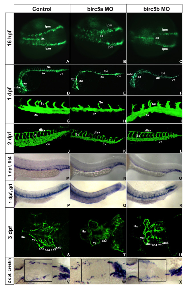
Fig. 3 Birc5 in vasculogenesis and angiogenesis. Tg(Fli:eGFP) (A-L) and Tg(Flk1:GFP) (S-U) embryos. A-C: 16 hpf (14 somites), Birc5a morphants with angioblast migration defects from lateral plate mesoderm (B) which are minor in Birc5b morphants (C). D-F: 1 dpf, Birc5a (E, H) and Birc5b (F, I) morphants have thinner axial vessels and poor caudal vein plexus development. G-I: 1 dpf Birc5a depletion (H) delays intersomitic vessels; not with Birc5b depletion (I). J-L: 2dpf Birc5a morphants with abnormal dorsal longitudinal anastomic and intersomitic vessels. Both morphants have poorly developed caudal vein plexus. M-O: flt4 at 1 dpf is reduced in posterior cardinal vein in both morphants (N, O). P-R: gridlock (grl) at 1 dpf is reduced with Birc5a depletion (Q), but not with Birc5b (R). S-U: 3 dpf Birc5a morphants have hypoplastic aortic arches (T). Birc5b depletion at 3 dpf causes hypoplasia of aortic arches 5–6 (U). V-X: Birc5a depletion decreases neural crest cells that migrate to branchial arches, detected with crestin probe. Birc5b depletion (L) reduces neural crest cells. ax: axial vessels, mhc: midbrain-hindbrain channel, Se: intersomitic vessels, aa: aortic arch, cv: caudal vein plexus, pcv: posterior cardinal vein, da: dorsal aorta, dlav, dorsal longitudinal anastomic vessel, Ha: hypobranchial artery, va: ventral aorta, lpm: lateral plate mesoderm.