
Fig. 4
- ID
- ZDB-IMAGE-090113-39
- Genes
- Publication
- Cerda et al., 2009 - RNA profiling of FAC-sorted neurons from the developing zebrafish spinal cord
- All Figures
- Figures for Cerda et al., 2009
|
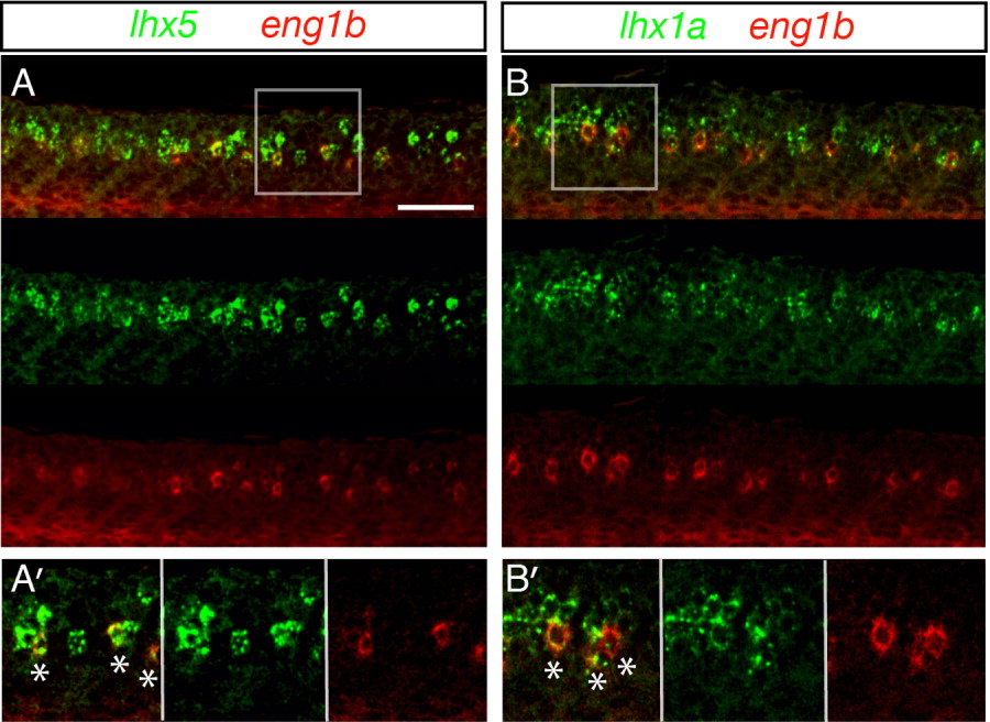
Fig. 4 Zebrafish V1 cells (CiAs) express lhx1a and lhx5. Confocal microscopy lateral views of fluorescent double in-situ hybridization of wildtype embryos at 27 hpf. In all cases, merged images and single channel views are shown. A: Z stack projections of lhx5 expression in green, eng1b expression in red. A′: Single focal plane of white box in A, showing lhx5 and eng1b co-localization. B: Z stack projections of lhx1a expression in green, eng1b expression in red. B′: Single focal plane of white box in B, showing lhx1a and eng1b co-localization. White stars in A′ and B′ merged images indicate double-labelled cells. Scale bar = 50 μm (A, B).