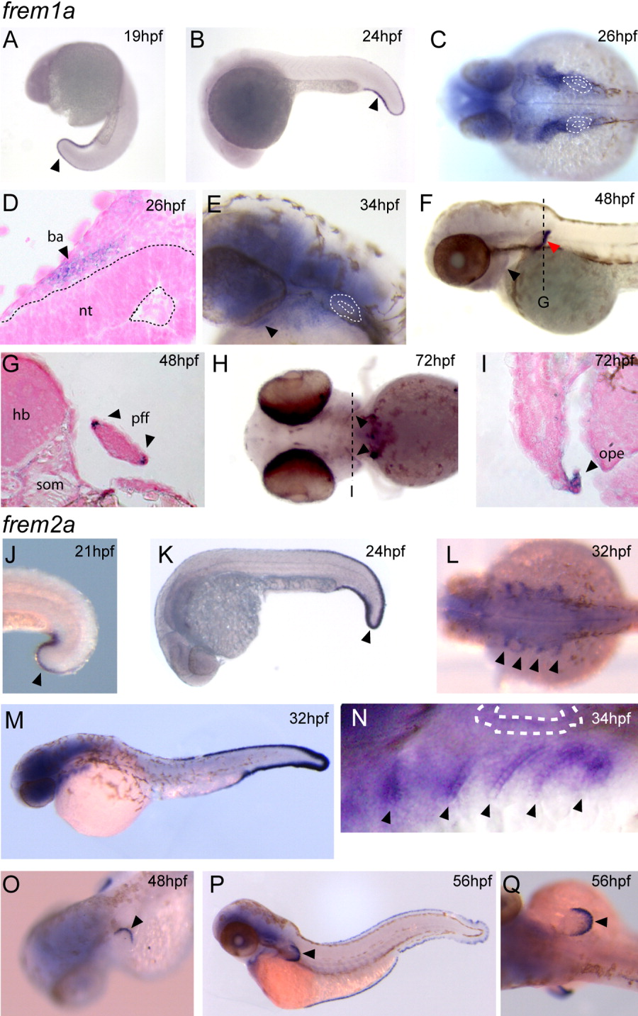
Fig. 2 Expression of frem1a and frem2a during development. Expression of frem1a (A-I) and frem2a (J-Q). A,B: Lateral views of frem1a expression detail expression of the gene in the developing caudal fin fold (arrowhead). C-E: At later stages the gene is also expressed in the developing branchial arches (C and D, in section; E, lateral view with hyoid indicated (arrowhead); otic vesicle highlighted in C and E). F-I: Expression is noted in the pectoral fins (F, lateral red arrowhead; G in section) and posterior ectodermal margin (PEM) of the hyoid arch (F and H, ventral, arrowhead) and in section (I, arrowhead). The line in H is the plane of section in I. J,K: Like frem1a, frem2a is expressed in the differentiating caudal fin fold (J and K, lateral views, arrowhead). L-N: Expression in the brachial arches initiates at 24 hours postfertilization (hpf) and by 32 hpf becomes strongest in the endodermal pouches (arrowheads in L, dorsal; in M, N dorsolateral, otic vesicle highlighted). O-Q: Expression is also detected in the pectoral fin folds (O-Q, arrowheads) and persists in the caudal fin folds of older fish (P, Q). ba, branchial arch; hb, hindbrain; som, somites; pff, pectoral fin fold; ope, operculum.
Image
Figure Caption
Figure Data
Acknowledgments
This image is the copyrighted work of the attributed author or publisher, and
ZFIN has permission only to display this image to its users.
Additional permissions should be obtained from the applicable author or publisher of the image.
Full text @ Dev. Dyn.

