
Fig. 3
|
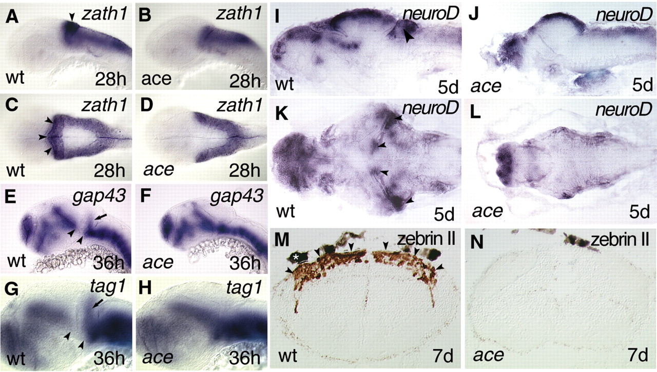
Fig. 3 Lack of cerebellar development in ace mutants. (A-H) Whole-mount in situ hybridisations. (I-L) In situ hybridisation of sections. (M-N) Immunohistochemistry of sections. (A-L) rostral to left; (A,B,E,H) lateral views; (C,D) dorsal views; (I,J) lateral views of sagittal sections; (K,L) dorsal views of horizontal whole brain sections; and (M,N) transversal hindbrain sections. zath1 expression is not detectable in the upper rhombic lips of ace mutants (B,D) in comparison with wild-type (A,C) embryos. Arrowheads (A,C) point to the upper rhombic lips expressing zath1 in wild-type embryos. No migrating granule cell precursors can be detected by analysing gap43 (E,F) and tag1 (G,H) expression in ace mutants. Arrows (E,G) mark the migrating granule cell precursors in the developing cerebellar anlage. Note that the ventral mesencephalic expression domain of both gap43 and tag1 are fused to the hindbrain expression domain. Arrowheads mark the gap between the rostral and caudal expression domains of gap43 (E) and tag1 (G). (I-L) Expression analysis of neurod fails to detect a cerebellar compartment containing granule cell precursors in the mutants. Arrowheads (I,K) point to the cerebellar anlage expressing neurod mRNA. (M,N) Immunohistochemical visualization of zebrin II, the evolutionarily conserved marker of Purkinje cells, fails to detect any of these cells in ace mutants. Arrowheads (M) point to the cerebellar plate loaded with zebrin II-expressing cells (brown staining). The white asterisk above the hindbrain section (M) marks a pigment granule.