
Fig. 5
|
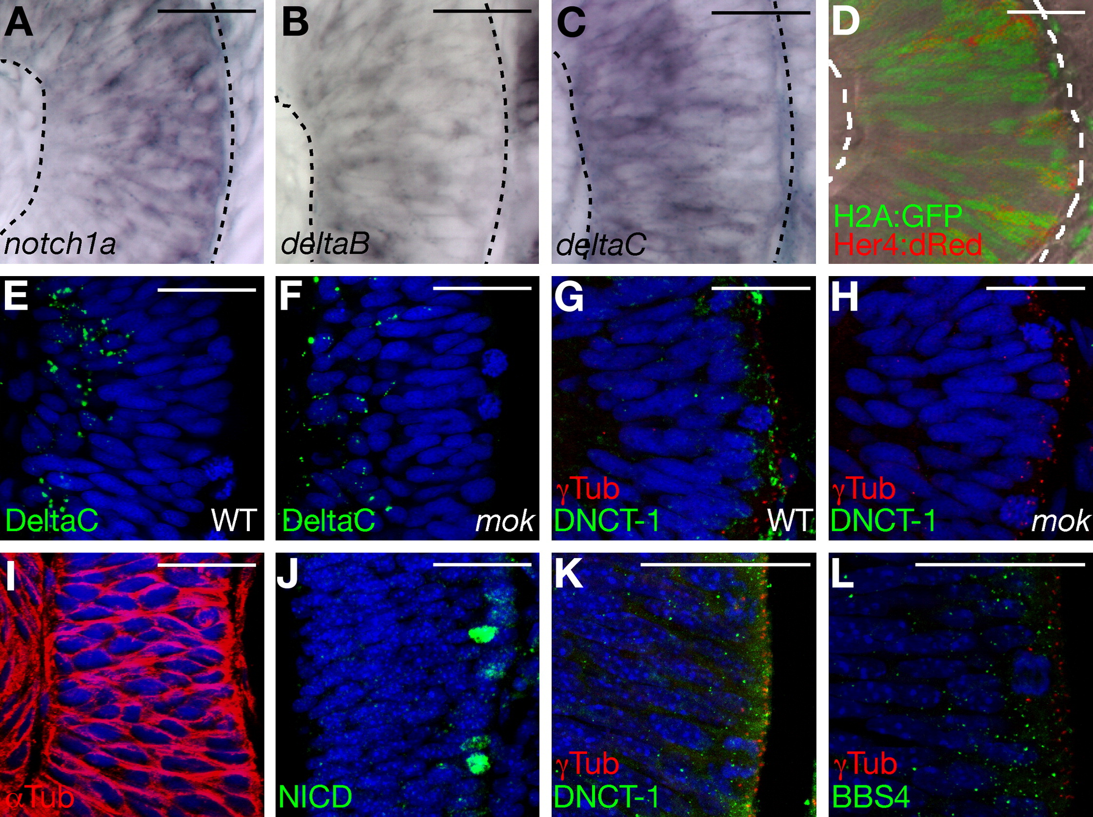
Fig. 5 A Gradient of Notch Signaling along the Apical-Basal Axis of the Developing Retina
(A–C) Coronal sections of 26 hpf retinas showing mRNA expression levels of components of the Notch/Delta signaling pathway. In situ hybridization shows higher levels of notch1a close to the apical surface of the retina (A) and deltaB and deltaC close to the basal surface (B and C).
(D) Optical section of a 33 hpf retina expressing her4:dRFP and H2A-GFP transgenes in a mosaic manner.
(E and F) Coronal sections of 26 hpf retinas stained with anti-DeltaC antibody, showing punctate cytoplasmic staining distributed in the basal half of the tissue both in wild-type and moks309 retinas.
(G and H) Coronal sections of 26 hpf retinas stained with anti-Dnct1 antibody, showing an enrichment at the apical surface in wild-type retinas (G), which is virtually absent in mutants (H).
(I) Coronal section of 26 hpf retina; α-tubulin staining reveals the parallel orientation of microtubules to the apical-basal axis.
(J–L) Sections of mouse retina. Activated Notch1 antibody labels a subset of nuclei at the apical surface (J). Anti-Dnct1 and anti-BBS4 antibodies show a cytoplasmic, punctated staining enriched at the apical surface. γ-tubulin staining (G, H, K, L) reveals the apical localization of the centrioles in retinal progenitors. In (D)–(L), DAPI (blue) stains the nuclei. In all panels, apical surface is on the left.
Scale bars, 25 μm (A–I) and 50 μm (J–L).
Reprinted from Cell, 134(6), Del Bene, F., Wehman, A.M., Link, B.A., and Baier, H., Regulation of neurogenesis by interkinetic nuclear migration through an apical-basal notch gradient, 1055-1065, Copyright (2008) with permission from Elsevier. Full text @ Cell