
Fig. 4
|
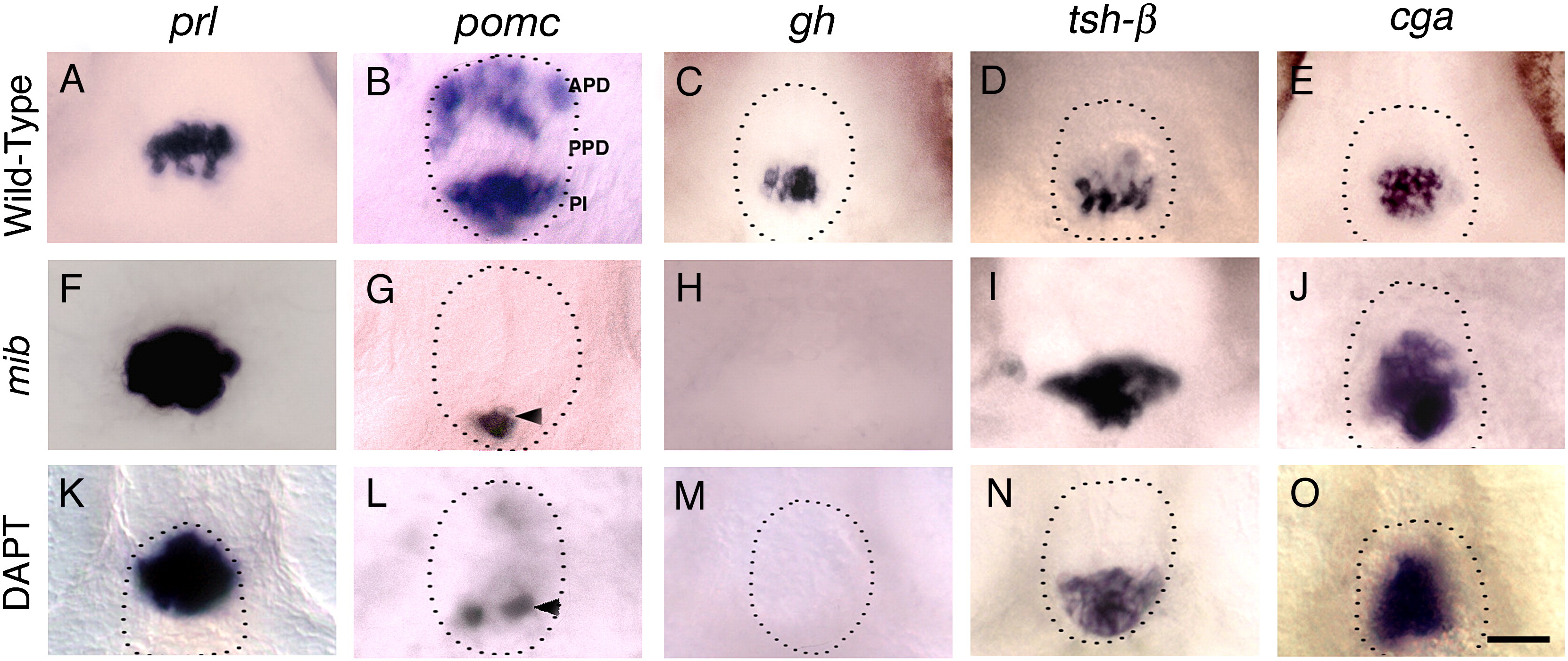
Fig. 4 Notch signaling is necessary for corticotrope and somatotrope differentiation. (A–E) Wild-type (Gene expression in DMSO treated control embryos, not shown, was same as in wild-type embryos in panels A–E), mib mutant (F–J) and DAPT treated embryos (K–O) labeled with probes for anterior pituitary hormones at 72 h. In mib mutant embryos, prl (F, n = 25/25), tsh-β (I, n = 23/23) and cga (J, n = 25/25) in the anterior pituitary is expanded, pomc expression is lost in APD and reduced in PPD (G, n = 26/26), and gh expression (H, n = 52/52) is completely lost compared to prl (A, n = 37), tsh-β (D, n = 40) and cga (E, n = 40), pomc (B, n = 20), gh expression (C, n = 40) in siblings. DAPT treatment between shield stage and 72 h leads to expanded prl (K, n = 26/33), tsh-β (n = 50/60) and cga expression (O, n = 35/42), whereas pomc expression in APD (L, n = 23/25) is lost or severely reduced, pomc expression in PI (L, n = 22/25) is somewhat reduced and gh expression (M, n = 35/40) is entirely absent in the PPD compared to wild-type embryos (A–E). Dotted lines indicate outline of anterior pituitary tissue (omitted in panels where pituitary border was ambiguous). Arrowhead shows pomc expression in PI. (A–O) Ventral views, anterior to the top. Scale bar: 25 μm (A–O).
Reprinted from Developmental Biology, 319(2), Dutta, S., Dietrich, J.E., Westerfield, M., and Varga, Z.M., Notch signaling regulates endocrine cell specification in the zebrafish anterior pituitary, 248-257, Copyright (2008) with permission from Elsevier. Full text @ Dev. Biol.