
Image
|
Figure Caption
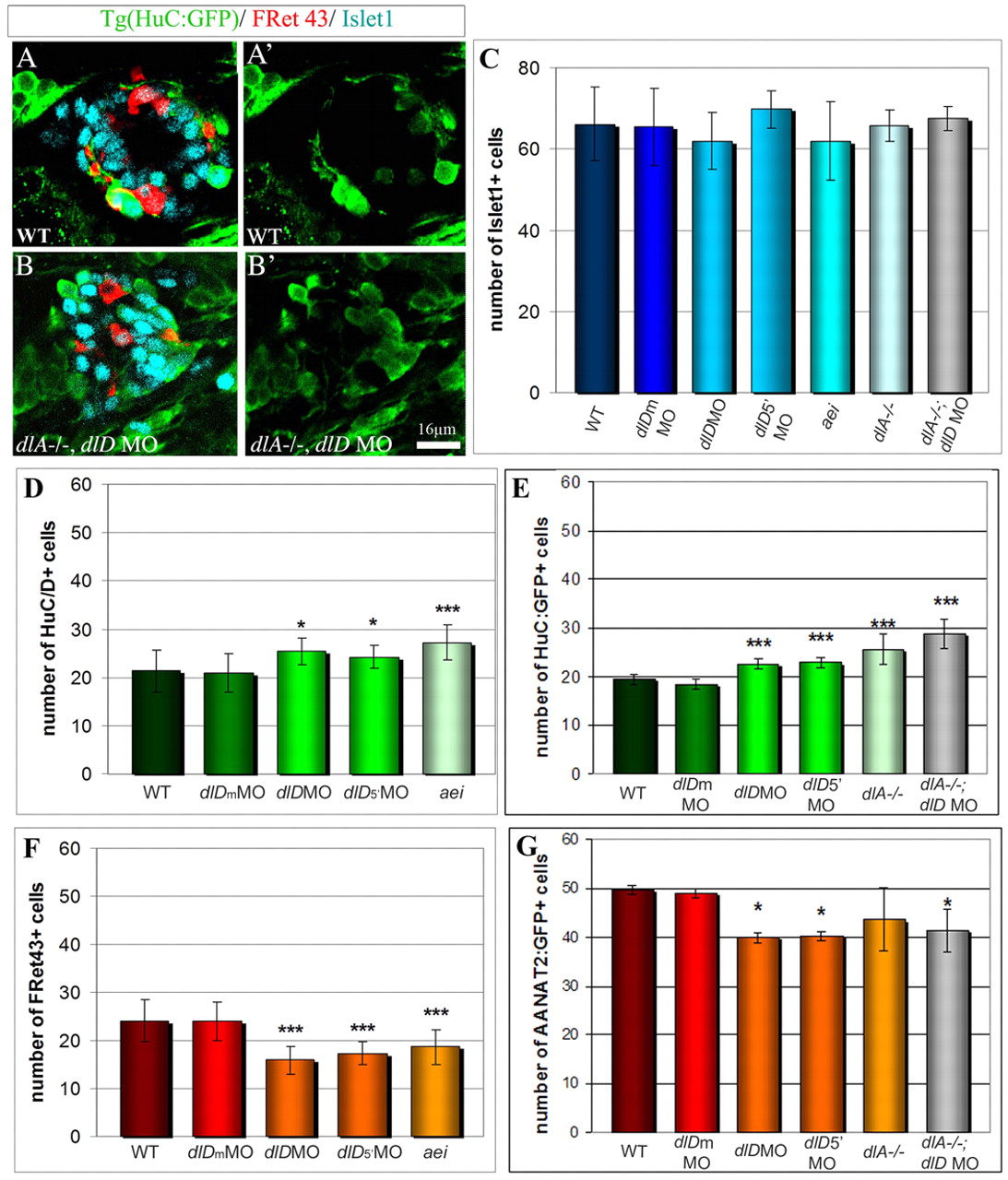
Fig. 5 Impaired photoreceptor/projection neurons ratio in embryos deficient for Delta genes. (A-B′) Confocal sections from wild-type (A-A′) and deltaA-/-, deltaD-morphant epiphysis (B-B′) showing GFP from Tg(HuC:GFP) (green), FRet43 (red) and Islet1 (purple) at 48 hours of development. (C-G)Average number of Islet1+ (C), HuC/D+ (D), Tg(HuC:GFP)+ (E), FRet43+ (F) and Tg(AANAT2:GFP)+ cells (G) in 48 hours embryos depleted for the function of deltaA and/or deltaD. Anterior is upwards. Scale bar: 16 μm. Error bars represent the standard deviation ***P<0.0005 using a t-test.
Figure Data
Acknowledgments
This image is the copyrighted work of the attributed author or publisher, and
ZFIN has permission only to display this image to its users.
Additional permissions should be obtained from the applicable author or publisher of the image.
Full text @ Development