
|
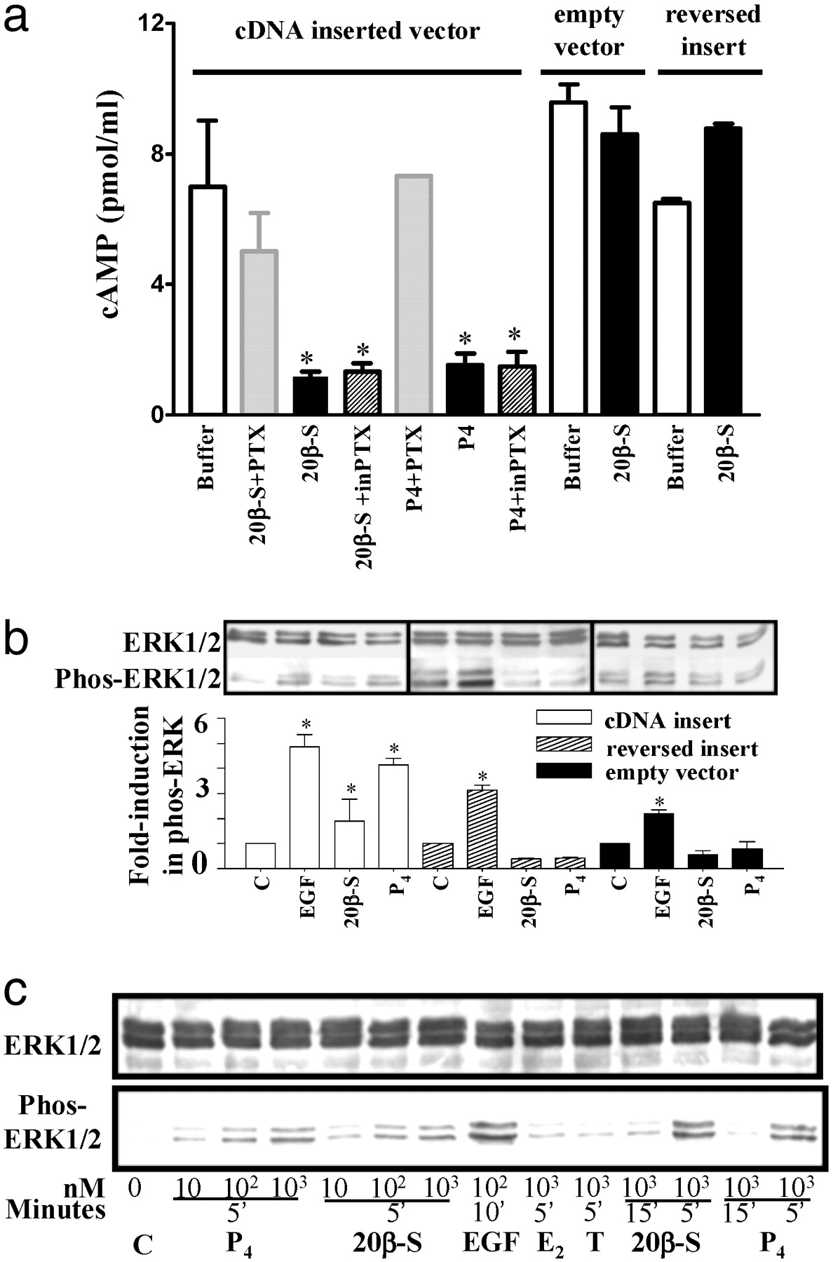
Fig. 4 Inhibition of cAMP production (a) and activation of MAP kinase signaling pathway (b and c) in response to progestin hormones in MDA-MB-231 cells stably transfected with putative mPR cDNA (means ± SEM, n = 4; *, P < 0.05). (a) Cells were preexposed to activated pertussis toxin (PTX) or inactive pertussis toxin (inPTX) for 30 min at 37°C before incubation with 20β-S or progesterone for 5 min. (b) MAP kinase activity (shown as increase in phospho-Erk1/2) in cells transfected with vector containing mPR insert (white bar), controls containing reversed insert (shaded bar) or vector alone (black bar) after a 5-min stimulation with 1 μM 20β-S or progesterone (P4). C, untreated control; EGF, human epidermal growth factor (1 μM, positive control). (c) Time- and dose-dependent activation of MAP kinase (gel loading: 20 μg per lane) in transfected cells treated with 20β-S, P4, estradiol-17β (E2), or testosterone (T).