
Image
|
Figure Caption
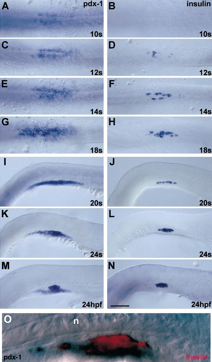
Fig. 3 Initiation of pdx-1 and insulin expression in the pancreatic primordium. (A–H) Dorsal and (I–N) lateral views of wild-type zebrafish embryos after in situ hybridization with pdx-1 (left panels) or insulin (right panels) antisense riboprobe at the (A, B) 10-somite stage; (C, D) 12-somite stage; (E, F) 14-somite stage; (G, H) 18-somite stage; (I, J) 20-somite stage; (K, L) 24-somite stage; and (M, N) 24 hpf. (O) 24 hpf embryo after in situ hybridization with both pdx-1 and insulin; n, notochord. The yolk was manually removed; anterior is to the left. Scale bar, 100 μm (N).
Figure Data
Acknowledgments
This image is the copyrighted work of the attributed author or publisher, and
ZFIN has permission only to display this image to its users.
Additional permissions should be obtained from the applicable author or publisher of the image.
Reprinted from Developmental Biology, 230(2), Biemar, F., Argenton, F., Schmidtke, R., Epperlein, S., Peers, B., and Driever, W., Pancreas development in zebrafish: early dispersed appearance of endocrine hormone expressing cells and their convergence to form the definitive islet, 189-203, Copyright (2001) with permission from Elsevier. Full text @ Dev. Biol.