
|
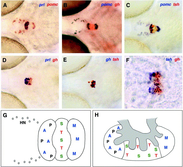
Fig. 5 Patterning of the zebrafish pituitary at 120 hpf. (A–F) Double in situ hybridizations, with probes indicated in top right corner in the corresponding colors. (A–E) Ventral view. (F) Sagittal section. (G, H) Schematic drawing of the pituitary composition in zebrafish larvae at 120 hpf (G; ventral view) and in adult teleost, modified from Norris (1997) (H; lateral view). A, corticotropes; M, melanotropes (both stained by pomc); P, lactotropes (prl); S, somatotropes (gh); T, thyrotropes (tsh); HN, endorphin-expressing hypothalamic neurons; in (H), the rudimentary neurohypophyis (infundibulum) is marked in gray.
Reprinted from Developmental Biology, 254(1), Herzog, W., Zeng, X., Lele, Z., Sonntag, C., Ting, J.W., Chang, C.Y., and Hammerschmidt, M., Adenohypophysis formation in the zebrafish and its dependence on sonic hedgehog, 36-49, Copyright (2003) with permission from Elsevier. Full text @ Dev. Biol.